Nikhil Prasad Fact checked by:Thailand Medical News Team Nov 08, 2026 18 minutes ago
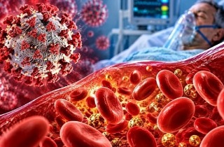
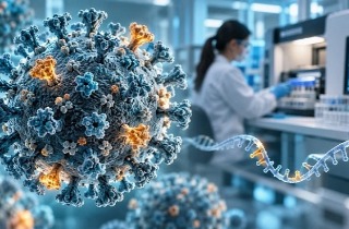
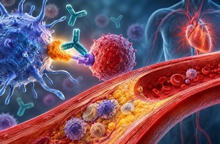
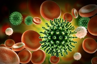
Scientists have discovered that exposure to SARS-CoV-2, the virus responsible for COVID-19, can cause significant disturbances within the body's immune tissues. The findings reveal that the virus can trigger excessive production of antibody-secreting cells while simultaneously reducing crucial T cells that protect against infections. Researchers also identified signs of inflammatory processes that could contribute to immune cell destruction.
 New research reveals how SARS-CoV-2 exposure can disrupt immune tissues, causing excessive
plasma cell development and inflammatory changes associated with the loss of protective T cells.
The study involved researchers from Third Military Medical University, the General Hospital of Western Theater Command, Dalian University College of Medicine, Chongqing Western Hospital, the People's Liberation Army Joint Logistic Support Force 989th Hospital, and the General Hospital of Central Theater Command, all in China.
Major Immune Cell Imbalances Discovered
Researchers initially examined lymph nodes collected from patients who died from COVID-19. Advanced genetic mapping revealed unusually large numbers of plasma cells, which manufacture antibodies, alongside severe losses of T cells. This imbalance is concerning because antibodies alone cannot provide complete immune protection. T cells are essential for eliminating infected cells and coordinating immune responses.
Experiments Reveal How SARS-CoV-2 Disrupts Immunity
To understand these changes, scientists created laboratory models of human tonsil tissue and exposed them to the original SARS-CoV-2 strain and Omicron XBB.1.5.
Both exposures promoted the transformation of B cells into antibody-producing plasma cells. However, researchers also detected heightened inflammatory activity among CD4 and CD8 T cells.
These cells produced increased amounts of TNF-alpha, interferon-gamma, and interleukin-2, substances that normally coordinate immune defenses but can contribute to tissue damage when inflammation becomes excessive.
Importantly, this
Thailand Medical News report highlights that these disturbances occurred even without detectable viral messenger RNA inside the experimental tissues.
Inflammatory Cell Death Signals Raise Concerns
Researchers additionally discovered increased activity involving NLRP3, caspase-8, and MLKL, molecules associated with PANoptosis, a form of inflammatory programmed cell death.
These findings suggest that excessive immune activation could contribute to T cell depletion, potentially compromising the body's ability to respond effectively to subsequent infections.
Notably, influenza exposure did not produce comparable changes in the tonsil models.
Conclusions
The findings indicate that SARS-CoV-2 exposure may disturb immune defenses through excessive plasma cell development, heightened inflammation, and activation of molecular pathways associated with immune cell death. Although fur
ther research is necessary to establish whether these mechanisms cause lasting immune damage, the results provide important clues about immune dysfunction during COVID-19.
The study findings were published as an abstract in the peer reviewed journal: Frontiers in Immunology.
https://www.frontiersin.org/journals/immunology/articles/10.3389/fimmu.2026.1959104/abstract
Read Also:
https://www.thailandmedical.news/articles/coronavirus
https://www.thailandmedical.news/articles/long-covid




 Share
 Tweet
 Share