Study Warns That the Peptide Endothelin-1 Drives Iron Imbalance and Joint Damage After COVID-19
Nikhil Prasad Fact checked by:Thailand Medical News Team Aug 16, 2026 1 month, 2 weeks, 5 days, 3 hours, 13 minutes ago
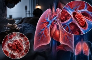
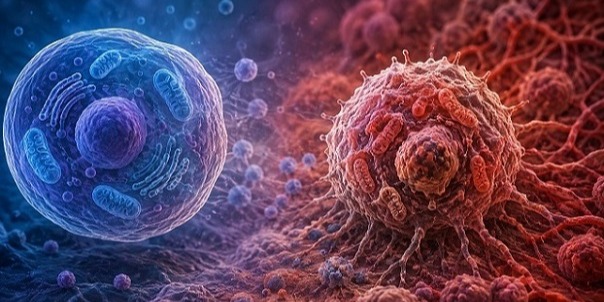
A new study has identified a troubling biological pathway that could explain why some people experience persistent joint, cartilage and bone problems after COVID-19. Researchers found that a small signaling peptide called endothelin-1 (ET-1) may help transmit damaging effects from SARS-CoV-2-injured lungs to distant skeletal tissues.
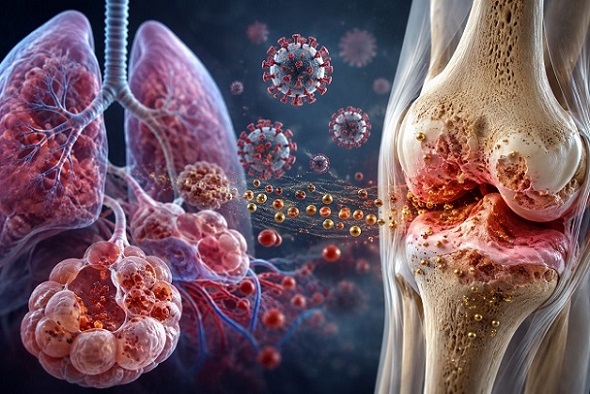 Researchers have identified endothelin-1 as a possible link between SARS-CoV-2 lung injury, abnormal
iron accumulation and subsequent cartilage and joint damage
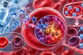
The findings point to a previously underappreciated lung-joint axis, in which increased ET-1 disrupts the body's normal handling of iron, resulting in abnormal iron accumulation, oxidative damage and the loss of important cartilage cells.
Researchers From International Institutions
The researchers were from The Hong Kong Polytechnic University; Guizhou Provincial Center for Disease Control and Prevention; The Hong Kong Polytechnic University Shenzhen Research Institute; The University of Hong Kong; Southern University of Science and Technology; Hong Kong Baptist University; West China Hospital of Sichuan University; Queensland University of Technology; The University of Hong Kong-Shenzhen Hospital; and the Centre for Virology, Vaccinology and Therapeutics at Hong Kong Science and Technology Park.
Lung Cells Produce More Endothelin-1 After Infection
The scientists found that SARS-CoV-2 infection increased ET-1 particularly in alveolar type II, or AT2, cells, which are important epithelial cells within the lungs.
Analysis of human lung samples showed that these cells displayed substantial increases in the gene responsible for producing ET-1. At the same time, several genes controlling iron uptake, storage and metabolism became abnormal.
The researchers subsequently found similar changes in cartilage. Samples from people with a history of COVID-19 showed increased ET-1, higher levels of the iron-uptake protein TFR1 and signs of oxidative damage.
Iron Accumulation Could Be Damaging Cartilage
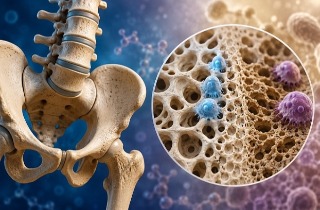
Experiments involving SARS-CoV-2-infected hamsters revealed cartilage cyst formation, thinning and disruption of growth plates, progressive cartilage-cell loss and deterioration of bone structure.
Importantly, lung ET-1 levels were strongly associated with TFR1 expression in cartilage. This suggests that abnormal signaling originating from infected or injured lungs could influence how cartilage cells handle iron.
Laboratory experiments provided additional evidence. When researchers genetically reduced ET-1 production in lung epithelial cells, spike-induced iron accumulation declined. Meanwhile, an iron-removing drug called deferiprone reduced abnormal iron deposits and partially restored cartilage-cell survival.
For readers of this Thailand
Medical News report, the findings suggest that excess iron is not simply an incidental feature following infection but could participate directly in damaging cartilage cells.
&l
t;strong>Endothelin Receptors May Also Interact with Viral Spike
Another major discovery involved the endothelin receptors ETAR and ETBR, which normally receive signals from ET-1.
Researchers found evidence that these receptors can also act as unconventional binding partners for the SARS-CoV-2 spike protein. Blocking or genetically reducing either receptor decreased spike uptake, while blocking both receptors produced the strongest suppression.
This creates a potentially damaging combination: SARS-CoV-2 can stimulate ET-1 production while ET-1 and its receptors promote abnormal iron handling and cellular injury.
Existing Drug Protected Joints in Infected Animals
The team therefore tested macitentan, an approved drug that blocks both endothelin receptors.
Early treatment of infected hamsters reduced viral levels in the lungs, pulmonary ET-1 expression, iron accumulation and markers of iron-associated cellular damage. It also improved cartilage structure, preserved cartilage cells and helped maintain growth-plate thickness.
Remarkably, delayed treatment between days 16 and 30 after infection still produced benefits. Macitentan reduced systemic iron accumulation and cartilage damage while increasing growth-plate thickness and cartilage-cell density. The treatment, however, did not completely reverse structural damage that had already developed.
Conclusions
The study provides important evidence that COVID-19-related skeletal problems could originate partly from biological signals generated in injured lungs. ET-1 appears capable of disturbing iron balance and promoting cartilage and bone damage, while endothelin receptors could represent future treatment targets. However, the human cartilage study involved relatively few samples, and the researchers stressed that larger prospective human studies and additional animal experiments are required before these findings can be translated into treatments for patients.
The study findings were published in the peer reviewed journal: Cell Reports.
https://www.sciencedirect.com/science/article/pii/S2211124726009198
Read Also:
https://www.thailandmedical.news/articles/coronavirus
https://www.thailandmedical.news/articles/long-covid




 Share
Share
 Tweet
Tweet
 Share
Share