Nikhil Prasad Fact checked by:Thailand Medical News Team Feb 05, 2026 4 months, 5 days, 6 hours, 53 minutes ago
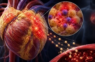
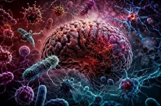
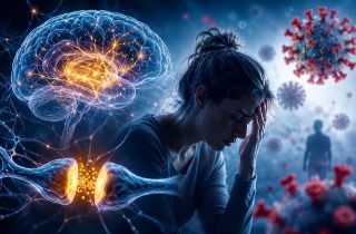
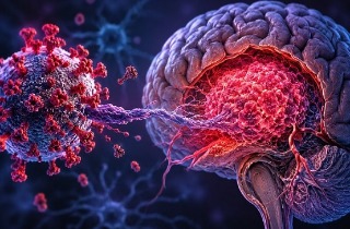
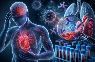
Medical News: Researchers are continuing to uncover why many individuals experience lingering neurological and psychological symptoms long after recovering from COVID-19. A new animal study now suggests that the SARS-CoV-2 spike protein alone, even without the live virus, may trigger slow and region-specific damage in the brain through a process known as ferroptosis, a form of iron-driven cell injury.
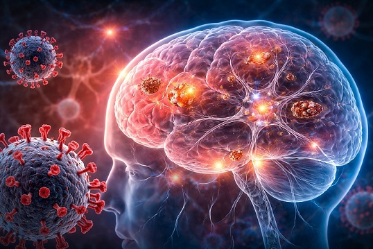 Spike protein exposure alone may quietly disrupt iron balance and trigger delayed brain cell injury
Ferroptosis is a recently discovered type of regulated cell death that occurs when iron metabolism becomes disrupted, antioxidant defenses fail, and damaging lipid oxidation builds up inside cells. This process has already been linked to aging, neurodegenerative diseases, and mood disorders, making it a strong candidate for explaining post-COVID neurological complications.
Why Researchers Focused on The Spike Protein
Instead of infecting animals with the full virus, which is lethal in this mouse model, scientists administered only the SARS-CoV-2 spike protein through the nasal passage. This approach mirrors real-world exposure routes and allows researchers to isolate the biological effects of the spike protein itself. This
Medical News report highlights how a single exposure produced measurable brain changes weeks later.
Different Brain Regions Showed Different Damage Patterns
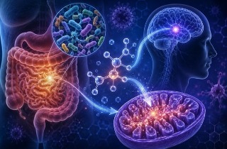
The study examined four key brain regions: the hippocampus, prefrontal cortex, cerebellum, and olfactory bulb. Each region showed a unique response over time.
In the hippocampus, which controls memory and learning, early changes were observed in proteins responsible for exporting iron from cells, suggesting an attempt to restore iron balance after disruption.
In the prefrontal cortex, which governs decision making and emotional regulation, early increases in iron import proteins were detected, raising concerns about iron overload and oxidative stress.
The cerebellum initially showed signs of protective antioxidant activation, but later developed markers of lipid damage, indicating delayed vulnerability.
The olfactory bulb showed late-stage iron transport changes, which may help explain why loss of smell persists in many long COVID patients.
Microscopic Evidence Confirms Cellular Injury
Using transmission electron microscopy, researchers directly observed structural damage in neurons from the hippocampus and prefrontal cortex. These changes included shrunken mitochondria, disrupted internal membranes, and weakened cell boundaries. Such features are considered hallmark signs of ferroptosis and indicate that the damage was not merely biochemical but structural.
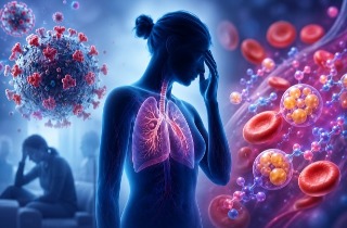
What These Findings Mean for Long COVID
The results suggest that the spike protein can initiate long-lasting stress responses in brain cells, even months after exposure. The time-dependent and reg
ion-specific nature of the damage mirrors the wide range of neurological symptoms reported by post-COVID patients, including brain fog, depression, memory loss, and sensory disturbances.
Study Limitations and Future Directions
The researchers caution that this was a preliminary study with a limited number of animals. They emphasize the need for larger and blinded studies, as well as investigations using ferroptosis-blocking drugs, to determine whether these findings translate directly to humans and whether intervention is possible.
Institutions Involved in The Research
The study was conducted by researchers from the Department of Neuroscience and the Department of Psychiatry and Psychology at the Mayo Clinic College of Medicine in Phoenix USA, the Department of Medical Physiology at Mansoura University and Mansoura National University in Egypt, and the Faculty of Dentistry at The Hashemite University in Jordan.
Conclusion
Taken together, the findings provide strong evidence that the SARS-CoV-2 spike protein alone can induce delayed and region-specific ferroptosis-related brain injury. These subtle but persistent cellular changes may contribute to long COVID neurological symptoms and underscore the importance of targeting iron metabolism and oxidative stress in future therapeutic strategies.
The study findings were published in the peer reviewed International Journal of Molecular Sciences.
https://www.mdpi.com/1422-0067/27/3/1526
For the latest COVID-19 news, keep on logging to Thailand
Medical News.
Read Also:
https://www.thailandmedical.news/articles/coronavirus
https://www.thailandmedical.news/articles/long-covid


 Share
Share
 Tweet
Tweet
 Share
Share