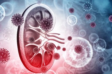
COVID-19 News: Study Finds That SARS-CoV-2 Nucleocapsid Protein Accumulates In Renal Tubular Epithelium Of Post-COVID-19 Patients, Possibility Causing AKI!
Nikhil Prasad Fact checked by:Thailand Medical News Team Nov 18, 2023 2 years, 2 months, 3 weeks, 5 days, 3 hours, 26 minutes ago
COVID-19 News: The lingering impact of the COVID-19 pandemic continues to unravel, transcending beyond the acute respiratory distress it initially manifested. Recent research conducted at Amsterdam University Medical Centre AMC in the Netherlands delves into the intricate interplay between the SARS-CoV-2 virus and renal complications in post-COVID-19 patients. The study, covered in this
COVID-19 News report, employing advanced techniques such as fluorescence microscopy and immunoelectron microscopy, sheds light on the accumulation of SARS-CoV-2 nucleocapsid protein in the renal tubular epithelium of patients who have survived and seemingly recovered from the virus.
 Understanding the Viral Presence in Kidney Tissue
In the wake of the COVID-19 pandemic, there has been a surge of interest in comprehending the long-term consequences of SARS-CoV-2 infection. While the primary focus of earlier research was on the respiratory and immune systems, the study aimed to investigate the presence of SARS-CoV-2 viral proteins and particles in the kidneys of both fatal COVID-19 cases and individuals experiencing kidney failure months after overcoming a SARS-CoV-2 infection.
The researchers utilized fluorescence microscopy to validate antibodies against various viral proteins, including nucleocapsid, membrane, and non-structural proteins. Intriguingly, the study revealed the accumulation of SARS-CoV-2 nucleocapsid (N) protein in the tubular epithelium of a patient experiencing acute kidney failure. Contrarily, other viral proteins and double-stranded RNA (dsRNA) associated with viral replication were not detected in these regions, suggesting a distinctive pattern of viral presence in the kidneys of these patients.
Insights into Post-COVID-19 Renal Complications
The importance of this study lies in its contribution to unraveling the enigmatic long-term effects of SARS-CoV-2 infection, particularly on the kidneys. Despite the evolving status of COVID-19 from a pandemic to a more conventional infectious disease, the intricate web of its aftermath continues to be a subject of intensive research. Acute kidney injury (AKI) has emerged as a significant extra-pulmonary complication of COVID-19, affecting a notable percentage of patients.
A meta-analysis indicated that approximately 4.5% of confirmed COVID-19 cases develop AKI, with higher incidence rates in critical cases. Recent studies have reported even higher rates, ranging from 15% to 25%, especially in COVID-19 patients with pre-existing health complications. Understanding the precise mechanisms through which SARS-CoV-2 affects the kidneys is crucial for enhancing treatment strategies during hospitalization and facilitating the recovery of post-COVID-19 patients grappling with kidney complications.
Unraveling the Origins of Acute Kidney Injury
The origins of acute kidney injury in COVID-19 patients are multifaceted, with several proposed causal factors. Hyper-coagulation, microangiopathy, rhabdomyolysis, endothelial activation, dysregulation of complement, and angiotensin-converting enzyme 2 (ACE2) pathway activation have all been suggested as potential contributors. Notably, the dire
ct cytotoxic effect of viral infection has also been postulated as a cause of kidney damage.
The kidney's susceptibility to SARS-CoV-2 infection is underscored by the high expression of the viral receptor ACE2 in both proximal tubular epithelial cells and parietal epithelial cells. However, the debate regarding whether the virus actively replicates in kidney tissue persists. Previous studies employing various techniques, including RNA in situ hybridization, immunohistochemistry, transmission electron microscopy, and quantitative reverse transcription PCR, have detected SARS-CoV-2 components in multiple extra-pulmonary tissues. Yet, the challenge lies in directly observing virus particles and replication organelles.
Visualizing Viral Proteins in Kidney Biopsies
The study at Amsterdam University Medical Centre sought to overcome this challenge by employing advanced microscopy techniques, aiming to visualize viral proteins in renal biopsies of COVID-19 patients. Initially, kidney samples from fatal COVID-19 cases were screened for viral proteins, but the results proved elusive, possibly due to the post-mortem collection conditions affecting tissue preservation.
However, a breakthrough came in the analysis of kidney biopsies from seven post-COVID-19 patients experiencing kidney problems. Using immunofluorescence microscopy, the researchers detected the accumulation of SARS-CoV-2 nucleocapsid (N) protein in tubular epithelial cells. This finding suggested that, even after apparent recovery from COVID-19, remnants of viral proteins may persist in the kidneys, raising questions about the potential implications for long-term kidney health.
Immunoelectron Microscopy Unveils Intricate Details
To delve deeper into the ultrastructure of the observed N protein accumulations, the researchers employed immunoelectron microscopy, a high-resolution technique that allows the precise localization of proteins on ultrathin sections.
The results revealed that the N protein accumulations were present in Golgi-like structures within the renal tubules, emphasizing the unexpected role of this protein in post-COVID-19 renal complications.
The localization of N protein on Golgi stacks was unexpected but not entirely unprecedented. Previous studies have indicated the fragmentation of the Golgi during early stages of SARS-CoV-2 infection in cultured cells. However, the overexpression of only the N protein did not induce similar rearrangements in the Golgi, setting the stage for further investigations into the cell biology mechanisms underlying this phenomenon.
The Dynamics of N Protein Accumulation
The researchers further explored the dynamics of N protein accumulation on Golgi stacks by determining its sub-Golgi localization. The majority of N protein was found on the cristae of Golgi-like structures, challenging conventional expectations of its subcellular distribution. The study also highlighted the scarcity of immunogold labeling on vesicles surrounding the Golgi stacks, raising questions about the nature of these vesicles and their potential role in virus replication.
Examining the Presence of dsRNA and Extracellular Vesicles
To ascertain whether renal epithelial cells with N protein accumulation produce viral RNA, the researchers tested for the presence of double-stranded RNA (dsRNA). While dsRNA was detected in SARS-CoV-2-infected Vero cells, its presence was not observed in renal biopsies with N protein accumulations, suggesting a lack of active viral replication in these kidney samples.
Additionally, the study explored the presence of extracellular vesicles in kidney tubule lumens, reminiscent of the lipid-filled compartments induced by the SARS-CoV-2 virus in lung tissue and Vero cells. Interestingly, a distal convoluted tubule was identified with electron-lucent compartments and vesicles resembling exosomes. This finding opens avenues for further investigations into the role of extracellular vesicles in the context of post-COVID-19 kidney complications.
Implications for Recurrence of Kidney Disease
The study's revelation of N protein accumulation in renal tubular epithelium, even in the absence of other viral proteins indicative of active replication, raises important considerations for the recurrence of kidney disease in post-COVID-19 patients. The N protein, being abundantly produced and remarkably stable, could potentially exert a prolonged influence on the immune system.
Previous research has demonstrated the N protein's interference in the innate immune response by modifying antiviral responses. Its presence in the cytosol has been shown to downregulate processing bodies, which are cytosolic RNA-protein granules involved in the regulation of inflammatory cytokine production.
Furthermore, the detection of N protein in urinary samples has been correlated with disease severity, emphasizing its significance beyond the immediate post-infection period.
Concluding Thoughts and Future Directions
In conclusion, the Amsterdam University Medical Centre's study offers a nuanced perspective on the renal implications of post-COVID-19 complications. The unexpected localization of SARS-CoV-2 N protein in Golgi-like structures within renal tubules opens up new avenues for understanding the dynamics of viral presence in different tissues.
As research in this field progresses, future investigations should focus on confirming the absence of active viral replication in human kidneys, delineating the specific mechanisms behind N protein accumulation, and exploring the potential long-term consequences on kidney health. This comprehensive analysis contributes to the evolving narrative of COVID-19's aftermath, urging continued exploration and understanding of the virus's intricate interactions with various organ systems.
The study findings were published in the peer reviewed journal: Microbiology Spectrum.
https://journals.asm.org/doi/10.1128/spectrum.03029-23
For the latest
COVID-19 News, keep on logging to Thailand Medical News.
Read Also:
https://www.thailandmedical.news/news/covid-19-news-insights-into-the-mechanism-behind-covid-19-induced-acute-kidney-injury-and-the-potential-therapeutic-role-of-quercetin
https://www.thailandmedical.news/news/covid-19-news-german-study-finds-that-urinary-n-terminal-pro-brain-natriuretic-peptide-predicts-acute-kidney-injury-and-severe-disease-in-covid-19
https://www.thailandmedical.news/news/japanese-study-finds-that-sars-cov-2-mediated-kidney-injury-can-be-prevented-by-inhibition-of-toll-like-receptor-4-and-interleukin-1-receptor
https://www.thailandmedical.news/news/breaking-news-fibrotic-events-in-kidneys-may-initiate-early-in-sars-cov-2-infection,-leading-to-pronounced-kidney-fibrosis-in-long-covid
https://www.thailandmedical.news/news/thailand-medical-news-warns-that-covid-19-infections-and-vaccinations-are-driving-exponential-incidences-of-acute-kidney-injury
https://www.thailandmedical.news/news/study-shockingly-shows-that-many-exposed-to-the-sars-cov-2-omicron-variant-exhibit-hematuria-and-proteinuria-early-signs-of-possible-kidney-damage
https://www.thailandmedical.news/news/covid-19-news-study-finds-that-non-omicron-variants-more-likely-to-cause-damage-to-the-kidney-s-filtration-system-than-omicron-sublineages
https://www.thailandmedical.news/news/breaking-covid-19-news-french-researchers-warn-that-post-covid-children-and-teenagers-are-at-risk-of-developing-kidney-disease
https://www.thailandmedical.news/news/breaking-covid-19-news-sars-cov-2-orf3a-protein-damages-renal-tubules-via-trim59-induced-stat3-activation-causing-acute-kidney-injury
https://www.thailandmedical.news/news/breaking-are-certain-newer-sars-cov-2-variants-causing-a-rise-in-chronic-kidney-disease-and-kidney-failure-in-countries-like-singapore-and-thailand
https://www.thailandmedical.news/news/breaking-u-s-cdc-reports-high-incidences-of-kidney-failure,-clots,-diabetes-and-heart-issues-in-post-covid-children-and-teenagers
https://www.thailandmedical.news/news/university-of-queensland-study-warns-that-millions-of-sars-cov-2-infected-individuals-are-not-aware-that-they-may-have-undiagnosed-acute-kidney-injury
https://www.thailandmedical.news/news/dutch-and-german-study-shows-that-sars-cov-2-directly-infects-the-kidneys-and-causes-tissue-scarring-which-ultimately-leads-to-kidney-damage-and-failu
https://www.thailandmedical.news/news/university-of-washington-study-confirms-that-sars-cov-2-can-directly-invade-human-kidney-cells-causing-a-range-of-kidney-issues-including-acute-kidney
https://www.thailandmedical.news/news/more-great-news-even-those-that-initially-had-mild-covid-19-symptoms-can-develop-kidney-disease-as-part-of-many-manifestations-of-long-covid
https://www.thailandmedical.news/news/good-news-study-finds-that-most-recovered-covid-19-patients-even-with-mild-infections-will-ultimately-develop-virus-induced-kidney-damage
https://www.thailandmedical.news/news/study-finds-that-covid-19-induced-acute-kidney-injury-similar-to-sepsis-caused-kidney-injury-and-that-mitochondrial-dysfunction-may-play-a-key-role
https://www.thailandmedical.news/news/covid-19-diagnostics-researchers-uncover-new-protein-biomarker-supar-to-identify-covid-19-patients-at-risk-of-acute-kidney-injury-aki
https://www.thailandmedical.news/news/warning-covid-19-latest-researchers-warn-of-epidemic-of-post-covid-19-kidney-disease-and-study-shows-many-will-die-or-never-recover-from-aki
https://www.thailandmedical.news/news/sars-cov-2-attacks-kidney-proximal-tubular-cells-causing-acute-fanconi-syndrome-according-to-french-study
https://www.thailandmedical.news/news/acute-kidney-injury-study-shows-that-acute-kidney-injury-(aki)-predominant-among-covid-19-patients
https://www.thailandmedical.news/news/covid-19-clinical-care-kidney-failure-emerging-as-a-common-occurrence-from-covid-19-infections
https://www.thailandmedical.news/news/breaking-more-emerging-chinese-research-studies-shows-that-the-sars-cov-2-coronavirus-also-attacks-the-kidneys,-pancreas-and-liver
