Nikhil Prasad Fact checked by:Thailand Medical News Team Oct 25, 2026 1 hour, 4 minutes ago
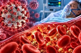
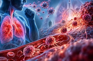
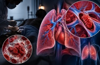
A new study has uncovered important changes in lung blood vessels among patients hospitalized with COVID-19 pneumonia, revealing how the distribution of different-sized vessels is closely associated with breathing difficulties and disease severity.
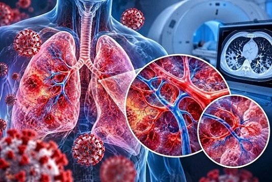 Chest CT analysis reveals distinctive lung blood vessel patterns associated with worsening oxygen
exchange and respiratory failure in COVID-19 pneumonia.
Researchers from the University of Pisa, University Hospital of Pisa, Pisa University-Hospital, and Mayo Clinic in the United States analyzed chest CT scans from 258 patients using specialized imaging software called CALIPER.
The findings suggest that examining blood vessel patterns could provide valuable information about how COVID-19 damages the lungs.
Examining Blood Vessels in COVID-19 Patients
The investigation involved patients hospitalized in Pisa, Italy, between March and April 2020. Their median age was 70 years, and 67% were men.
Using CALIPER software, researchers measured vascular-related structures across five categories, ranging from cross-sectional areas of 5 square millimeters or less to areas exceeding 20 square millimeters.
This
Thailand Medical News report highlights how these measurements revealed distinct relationships between vessel size, oxygen exchange, and respiratory support requirements.
Larger Vessel Proportions Linked to Severe Disease
The strongest finding involved differences between smaller and larger vascular structures.
Patients with greater proportions of structures measuring 20 square millimeters or less generally experienced milder illness and required less intensive breathing assistance.
Conversely, patients with greater proportions of structures exceeding 20 square millimeters were more likely to require advanced respiratory support.
Among the 258 patients, 32 required no supplemental oxygen, 138 received oxygen therapy, and 81 required noninvasive or invasive ventilation. Respiratory support information was unavailable for seven patients.
Overall, 52 patients died during hospitalization, representing approximately 20% of the study population.
Blood Vessel Patterns Reflect Oxygen Problems
Researchers also examined oxygen exchange using the PaO2/FiO2 ratio, which measures how effectively oxygen enters the bloodstream.
Higher proportions of smaller vascular structures were associated with better oxygen exchange, particularly among patients with ratios of 300 or higher.
Larger vascular structures showed the opposite relationship, becoming proportionally more prominent as oxygen exchange deteriorated.
These findings may reflect inflammation, vascular injury, altered blood circulation, and increased resistance within pulmonary vessels.
However, the measurements represent relative vascular proportions rather than direct evidenc
e that individual vessels disappeared or enlarged.
Conclusions and Future Implications
The findings suggest that quantitative CT analysis could improve understanding of vascular abnormalities accompanying severe COVID-19 pneumonia and potentially support future research into pulmonary hypertension.
Nevertheless, this retrospective, single-center study cannot establish causation or demonstrate predictive accuracy. Larger investigations are needed to validate these associations and determine their clinical usefulness.
The study findings were published in the peer-reviewed journal: Methods and Protocols.
https://www.mdpi.com/2409-9279/9/6/157
Read Also:
https://www.thailandmedical.news/articles/coronavirus
https://www.thailandmedical.news/articles/long-covid 



 Share
 Tweet
 Share