Nikhil Prasad Fact checked by:Thailand Medical News Team Feb 26, 2026 3 months, 5 days, 20 hours, 30 minutes ago
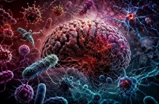
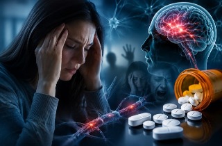
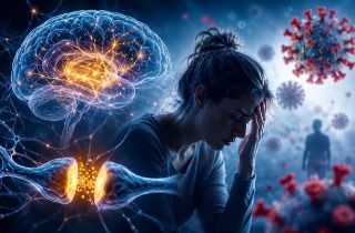
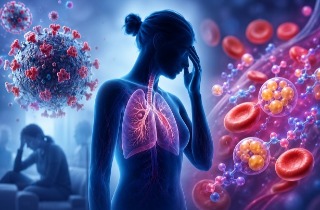
Medical News: Sepsis, a life-threatening reaction to infection, does not only attack the lungs, kidneys, or heart. It can also strike the brain, causing confusion, delirium, memory loss, and even coma. Now, scientists have uncovered new evidence that tiny proteins called histones may play a central role in damaging the brain during severe sepsis.
 Extracellular histones infiltrate the brain during sepsis, break down its protective barrier, and ignite
damaging inflammation
Researchers from the Division of Allergy and Clinical Immunology, Department of Internal Medicine, the Department of Pathology, and the Department of Surgery at the University of Michigan Medical School in Ann Arbor, Michigan, have discovered how these proteins escape into the bloodstream and breach the brain’s protective shield, triggering widespread inflammation.
How Sepsis Turns the Brain Against Itself
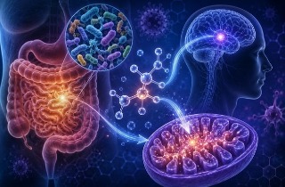
Histones normally live safely inside the nucleus of cells, where they help package DNA. But during sepsis, massive immune activation and cell damage cause histones to spill into the bloodstream. This
Medical News report highlights how these escaped histones can become toxic.
Using a well-established mouse model of polymicrobial sepsis, the team found that histones began appearing in brain tissue as early as four hours after sepsis onset. By eight hours, levels had surged dramatically, reaching their peak between eight and twelve hours before gradually declining.
Under normal conditions, the brain is protected by the blood–brain barrier, a tightly sealed layer of cells that prevents harmful substances from entering. However, the researchers demonstrated that this barrier became leaky during sepsis. Small tracer molecules were able to pass into the brain within eight hours, and larger molecules followed by 24 hours, showing that the damage worsened over time.
A Chain Reaction of Inflammation
The study revealed that histone accumulation in the brain coincided with activation of the complement system, particularly a powerful inflammatory pathway involving the molecule C5a and its receptor. Expression of the C5a receptor in brain tissue increased up to eightfold during early sepsis.
Once histones entered the brain, they did not remain passive. The team found that astrocytes and microglia, the brain’s main support and immune cells, absorbed these histones. Under the microscope, histones were clearly visible inside these cells, especially 18 hours after sepsis began.
Astrocytes reacted strongly. Levels of GFAP, a marker of astrocyte activation, rose significantly within four hours and remained elevated. Other markers of glial activation, including S100B and Iba1, also increased, confirming that both astrocytes and microglia were highly stimulated.
In laboratory experiments, astrocytes exposed to histones released large amounts of inflammatory cytokines such as IL-6 and TNF-alpha. Histones also triggered rapid calcium surges inside astrocytes within
seconds, a clear sign of cellular activation. Most strikingly, histones activated the NLRP3 inflammasome, a molecular alarm system that amplifies inflammation. IL-1β gene expression increased hundreds of times under inflammasome stimulation conditions.
A Dangerous Feedback Loop
The findings suggest a vicious cycle. Sepsis triggers complement activation and immune cell overdrive. Neutrophils release extracellular traps containing histones. The blood–brain barrier becomes compromised, allowing histones to enter the brain. Once inside, histones activate astrocytes and microglia, which release more inflammatory signals, further weakening the barrier and worsening brain injury.
Importantly, astrocytes were shown not only to absorb histones but also to release them when stimulated by inflammatory molecules such as C5a, LPS, or PMA. This means brain cells themselves may help sustain the inflammatory storm.
What This Means for Patients
Sepsis-associated encephalopathy affects a large percentage of critically ill patients and is linked to long-term cognitive problems. Yet there are no specific treatments targeting the brain complications of sepsis.
The researchers conclude that extracellular histones and complement pathways may represent promising therapeutic targets. Neutralizing histones or blocking complement activation could potentially protect the blood–brain barrier, reduce inflammation, and limit lasting brain damage.
In conclusion, this study provides compelling evidence that extracellular histones are not merely byproducts of infection but active drivers of brain injury in sepsis. By breaching the blood–brain barrier, activating complement pathways, and triggering powerful inflammatory responses in astrocytes and microglia, histones create a self-amplifying cycle of neuroinflammation. Targeting these molecules could open the door to new strategies aimed at preventing cognitive decline and improving survival outcomes in patients suffering from severe sepsis.
The study findings were published in the peer reviewed International Journal of Molecular Sciences.
https://www.mdpi.com/1422-0067/27/5/2126
For the latest on Sepsis, keep on logging to Thailand
Medical News.
Read Also:
https://www.thailandmedical.news/articles/sepsis


 Share
Share
 Tweet
Tweet
 Share
Share