Study Uncovers Lasting Liver Damage After COVID-19 Especially in Overweight Individuals
Nikhil Prasad Fact checked by:Thailand Medical News Team Oct 18, 2026 44 minutes ago
COVID-19 may leave behind more than respiratory damage, particularly in individuals with obesity. New research suggests that SARS-CoV-2 infection can trigger persistent inflammation, metabolic disturbances, and cellular changes in the liver, even after the virus has been cleared.
 New research reveals how obesity may prolong liver inflammation, fat accumulation, and metabolic
abnormalities after COVID-19 infection
Researchers from the Beirne B. Carter Center for Immunology Research, Department of Microbiology, Immunology and Cancer Biology, and Division of Infectious Diseases and International Health, Department of Medicine, all at the University of Virginia, United States, uncovered these changes in an experimental mouse study.
The findings raise concerns about how obesity may complicate recovery from COVID-19 and potentially contribute to prolonged liver dysfunction.
Investigating Liver Damage After COVID-19
For this
Thailand Medical News report, researchers compared lean mice with mice that developed obesity through high-fat diets.
Both groups were infected with a mouse-adapted SARS-CoV-2 strain. Scientists examined liver tissues before infection and at seven and 28 days afterward.
Using single-cell RNA sequencing, they investigated how individual liver cells responded to infection and whether their biological activities returned to normal during recovery.
Liver Injury Persists After Viral Clearance
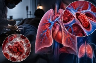
Obese mice experienced greater illness severity, reduced survival, and more extensive lung damage.
Although detectable virus disappeared by day 28, obese mice continued showing lung inflammation, collagen accumulation, and tissue changes associated with fibrosis.
Liver examinations revealed similarly concerning abnormalities.
Obese mice accumulated significantly more fat droplets within liver tissue during acute infection. Excessive fat accumulation remained evident 28 days later.
Researchers also detected increased apoptosis, a form of programmed cell death, indicating continued liver-cell injury.
Blood testing revealed elevated glucose and cholesterol levels in obese mice. Alanine aminotransferase, an enzyme indicating liver-cell damage, increased significantly during acute infection at day seven.
Importantly, some metabolic abnormalities were already associated with obesity before infection.
Immune Cells Remain Abnormally Activated
One striking discovery involved macrophages, immune cells responsible for clearing damaged tissue and regulating inflammation.
During acute infection, obese mice developed increased liver infiltration by neutrophils and inflammatory monocytes.
Although these populations subsequently declined, certain macrophage populations remained unusually abundant at day 28.
These included lipid-associated m
acrophages and inflammatory monocyte-derived macrophages.
Gene-expression analysis revealed persistent activation of inflammatory pathways involving IL6-JAK-STAT3, TNF-NF-kappaB, and complement signaling.
Kupffer cells, specialized liver macrophages, also demonstrated altered metabolic activity, reduced fatty acid oxidation, and weakened insulin-response pathways.
These findings suggest that obesity may interfere with the liver's ability to resolve inflammation following infection.
Metabolism and Blood Vessels Also Change
Hepatocytes, the liver's principal functional cells, showed increased gene signatures associated with glucose production and fatty acid uptake.
At day seven, researchers observed increased fat-production activity and reduced fat-breakdown signatures.
By day 28, hepatocytes continued displaying abnormalities involving fat metabolism, collagen production, and inflammatory signaling.
Blood-vessel lining cells also remained altered.
Unlike lean mice, whose endothelial cells showed recovery-related vascular programs, obese mice displayed persistent immune activation and increased expression of genes associated with platelet aggregation and blood clotting.
Computational analysis additionally suggested altered communication between macrophages and hepatocytes, potentially connecting persistent inflammation with disrupted metabolism.
Conclusions and Implications
The findings suggest that obesity can prolong liver inflammation and metabolic disruption after SARS-CoV-2 infection, potentially increasing vulnerability to post-COVID complications. However, the experiments involved only male mice, and pooled sequencing samples limited biological replication. Human studies are essential to determine whether these abnormalities persist long-term and contribute directly to liver disease.
The study findings were published in the peer reviewed journal: Frontiers in Immunology.
https://www.frontiersin.org/journals/immunology/articles/10.3389/fimmu.2026.1910050/full
Read Also:
https://www.thailandmedical.news/articles/coronavirus
https://www.thailandmedical.news/articles/long-covid




 Share
Share
 Tweet
Tweet
 Share
Share