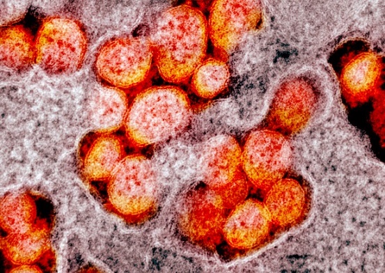
SARS-CoV-2 E Protein Found to Create Localized Membrane Deformations in Host Cells!
Nikhil Prasad Fact checked by:Thailand Medical News Team May 18, 2025 9 months, 6 days, 23 hours, 45 minutes ago
SARS-CoV-2 E Protein and Its True Role in Membrane Bending
Researchers from the Center for Computational and Integrative Biology (CCIB) at Rutgers-Camden and the Department of Physics at Rutgers-Camden, new Jersey-USA have uncovered new insights into the behavior of the SARS-CoV envelope (E) protein of both the SARS-CoV-1 and SARS-Co-2 viruses and how it interacts with cellular membranes. For years, scientists speculated that the E protein, a tiny but critical structural part of the coronavirus, might be responsible for bending membranes and helping the virus bud and shape itself. However, new findings challenge these assumptions and offer a deeper look into the true nature of this viral protein.
 SARS-CoV-2 E Protein Found to Create Localized Membrane Deformations in Host Cells!
The E protein has long been thought to play a pivotal role in the viral budding process by causing long-range bending of membranes. This
Medical News report shows that while the E protein does indeed distort the membranes, the deformations are very localized and do not stretch across large membrane areas as previously believed. The findings help clarify a lot of confusion caused by conflicting earlier studies and reveal that past results may have been influenced by hidden technical issues during computer simulations.
A Key Structural Component of the Virus
The E protein is one of the smallest but most important parts of the coronavirus structure. Despite its size, it is essential for the virus to replicate and exit infected cells. During the COVID-19 pandemic, the E protein remained highly stable with very few mutations, making it an attractive target for drug development.
The structure of the E protein includes three parts: a short N-terminal domain (NTD), a transmembrane domain (TMD), and a C-terminal domain (CTD). It usually forms groups of five called pentamers that embed themselves into cellular membranes. Scientists believed that these pentamers could bend the cell membrane to assist in virus formation.
Uncovering Simulation Artifacts That Misled Past Research
In the past, some studies had reported that the E protein could cause dramatic, long-range bending of cell membranes. However, the Rutgers-Camden team discovered that such results might have been caused by an unnoticed simulation error called a "barostat artifact."
Using advanced simulations that corrected these errors, the researchers found that when the E protein is properly simulated, it causes only severe local bending near itself but does not deform the entire membrane. Essentially, the membrane returns to its normal shape just a few nanometers away from the protein.
The Hidden Role of Hydrophobic Mismatch
One of the major discoveries of the study was the effect of "hydrophobic mismatch." This term describes the difference in thickness between the oily middle parts of the cell membrane and the oily parts of the protein that interact with it. When the thickness does not match, it causes the membrane to compress or stretch to adjust.
/>
The researchers found that hydrophobic mismatch plays a critical role in the local bending caused by the E protein. When the mismatch is large, the membrane around the E protein deforms more significantly. However, even in the worst cases, these deformations stay localized and do not extend across the membrane.
Asymmetry Matters More Than We Thought
Interestingly, the team also discovered that the membrane does not bend symmetrically around the E protein. Because of the E protein's odd shape, it causes one side of the membrane to be compressed more than the other. This creates an "asymmetry" that helps the membrane lower the energy cost of bending.
The study found a strong correlation between this asymmetry and the membrane's curvature. In simple terms, when one side of the membrane becomes thinner, it encourages bending toward that side. This coupling of thickness and curvature might help the virus optimize the shape of its envelope during formation, although it does not seem enough on its own to drive full virus budding.
Testing Different Membrane Types
To ensure their findings were not limited to a specific type of membrane, the researchers tested the E protein in various artificial membranes with different thicknesses and lipid flexibilities. In all cases, the same pattern emerged: severe local bending, but no large-scale deformation.
They also tested membranes made of "stiffer" lipids versus "more flexible" lipids. They found that more flexible membranes (those with unsaturated fats) could better accommodate the protein without bending too much, while stiffer membranes bent more around the E protein.
Concluding Remarks
The findings of this study offer important corrections to previous assumptions about the SARS-CoV-E protein. The results clearly show that, contrary to some earlier reports, the E protein by itself does not induce large-scale bending of cell membranes. Instead, it causes significant but very localized deformations, primarily influenced by hydrophobic mismatch and asymmetry between the membrane layers. These localized changes may contribute to virus formation when many E proteins or other viral proteins work together but are insufficient alone.
Importantly, the research highlights how even small technical issues in simulations can lead to major misunderstandings in science. By carefully controlling for artifacts, the Rutgers-Camden researchers have provided a more accurate picture of the E protein's role.
These insights could prove invaluable for future drug development targeting the E protein. By understanding exactly how the E protein affects membranes, scientists can better design therapies that disrupt the virus's ability to assemble and spread.
Overall, while the E protein remains a critical player in the viral life cycle, its true power lies not in overwhelming membrane bending but in precise, localized shaping of its environment a small but vital piece of the viral assembly puzzle. This research paves the way for more sophisticated strategies to combat current and future coronaviruses.
The study findings were published on a preprint server and are currently being peer-reviewed.
https://www.biorxiv.org/content/10.1101/2025.04.18.649534v1
For the latest COVID-19 News, keep on logging to Thailand Medical News.
Read Also:
https://www.thailandmedical.news/news/sars-cov-2-envelope-protein-quietly-destroys-human-cell-health
https://www.thailandmedical.news/news/french-scientists-discover-that-sars-cov-2-envelope-protein-s-pdz-binding-motif-disrupts-host-s-epithelial-cell-cell-junction
https://www.thailandmedical.news/news/sars-cov-2-e-protein-triggers-neuron-cell-death-via-calcium-release
https://www.thailandmedical.news/articles/coronavirus
https://www.thailandmedical.news/pages/thailand_doctors_listings
