Nikhil Prasad Fact checked by:Thailand Medical News Team Sep 16, 2026 1 day, 1 hour, 48 minutes ago
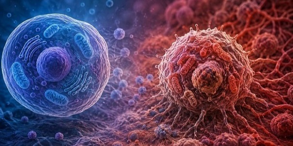
Glaucoma is usually associated with damaging pressure inside the eye, but a new scientific review suggests the disease may involve a more complex chain of neurodegenerative and immune changes. Researchers examined evidence linking abnormal tau protein, altered microglia—the nervous system’s resident immune cells—and damage to retinal ganglion cells (RGCs), whose axons form the optic nerve.
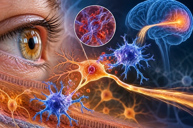 New evidence suggests abnormal tau and remodeled immune cells may interact in glaucoma, potentially contributing
to retinal and optic-nerve damage
The review was conducted by Chengdong Li, Ruijia Song, and Ling Yu from the Department of Ophthalmology, The Affiliated Hospital of Southwest Medical University, Southwest Medical University, Luzhou, Sichuan, China.
Evidence Points Beyond Eye Pressure
For this Thailand
Medical News report, the researchers searched PubMed from its inception through May 2026, drawing together human studies, experimental glaucoma and ocular-hypertension research, optic-nerve injury studies, and relevant tauopathy evidence.
The evidence indicates that glaucoma can affect more than the retina and optic nerve. Changes have also been reported farther along the visual pathway. In glaucoma, tau—a protein important for stabilizing neuronal microtubules and supporting axonal structure—shows altered abundance, phosphorylation, and location.
Human glaucomatous retina has shown reduced BT2 tau immunoreactivity and increased AT8 staining, although the AT8 signal was predominantly found in retinal horizontal cells. In a rat ocular-hypertension model, researchers detected tau accumulation, altered phosphorylation, movement into somatodendritic compartments, and T22-reactive tau oligomers. Importantly, oligomeric tau evidence in glaucoma remains limited to that model.
Tau Manipulation Changes Retinal Injury
Experimental findings suggest tau may be more than a marker of damage. Tau-targeted siRNA reduced both cell-body and axonal RGC injury. AAV9-mediated tau silencing also reduced inner-retinal degeneration and strengthened Akt/Erk survival signaling, while tau overexpression worsened retinal injury.
At the same time, glaucoma-associated microglia lose normal homeostatic programs and acquire neurodegeneration-related characteristics involving APOE, Galectin-3, complement activity, inflammation, and phagocytosis-related pathways. Complement is especially notable because inhibiting its classical pathway preserved early dendritic and synaptic architecture in experimental glaucoma.
The review proposes a cautious model: tau-related stress could make dendrites, synapses, or axons more vulnerable to recognition, while remodeled microglia and complement pathways could influence which damaged structures are cleared. However, researchers emphasized that glaucoma studies have not demonstrated that tau directly triggers microglia to engulf viable RGC structures.
Biomarkers Could Offer a Clinical Window
Aqueous humor may provide measura
ble clues. Total tau, phosphorylated tau, neurofilament light chain, GFAP, APOE, Galectin-3, complement-related molecules, and inflammatory markers could potentially reveal simultaneous neuronal and immune injury. Yet these biomarkers cannot establish which process begins first or prove causation.
Conclusion
The evidence supports tau disruption, immune-cell remodeling, and complement-linked synaptic vulnerability as important features of glaucoma, but their causal relationship remains unproven. Future experiments must track tau species, microglial states, targeted engulfment, axonal function, synaptic integrity, and RGC survival together over time to determine whether these processes truly form a damaging biological chain.
The study findings were published in the peer reviewed journal: Frontiers in Neurology.
https://www.frontiersin.org/journals/neurology/articles/10.3389/fneur.2026.1940010/full
Read Also:
https://www.thailandmedical.news/articles/glaucoma-news




 Share
Share
 Tweet
Tweet
 Share
Share