BREAKING NEWS
For the latest on Thailand Medical Industry, Thailand Doctors, Thailand Medical Research, Thailand Hospitals, Thailand Wellness Initiatives and the latest Medical News
 Share
Share
 Tweet
Tweet
 Share
Share
Medical News: Although COVID-19 is best known as a respiratory disease, scientists are continuing to uncover ways the virus can affect other organs long after infection begins. A new study has revealed that even mild COVID-19 infections can disrupt liver health through a surprising biological pathway that allows viral proteins produced in the lungs to travel to the liver and interfere with normal fat metabolism.
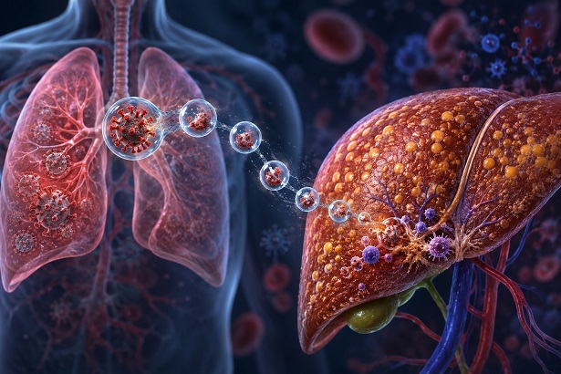
Scientists discover that a SARS-CoV-2 protein can travel from the lungs to the liver, causing fat buildup, inflammation,
and early fibrosis even during mild COVID-19
Researchers found that a SARS-CoV-2 protein known as ORF3a can be transported from the lungs to the liver inside tiny particles called exosomes. Once it reaches the liver, the protein promotes fat accumulation, inflammation, and early signs of fibrosis, even when the virus itself is not present in liver tissue.
Liver Problems Common Even in Mild COVID-19
The study began by examining 133 patients with mild COVID-19 and comparing them with 63 individuals who tested negative for SARS-CoV-2. None of the participants had pre-existing liver disease.
The findings were concerning. More than half of the patients with mild COVID-19 showed abnormal liver function tests. Elevated levels of liver enzymes such as ALT, AST, and GGT were commonly observed, indicating that liver stress and injury can occur even in cases that are not severe enough to require intensive medical care.
These observations raised an important question: if the virus remains largely confined to the respiratory tract during mild infections, how does the liver become affected?
A Viral Protein Travels from the Lung to the Liver
To answer this question, researchers used a mouse model of mild COVID-19. While active viral replication was detected only in the lungs, scientists discovered that the viral protein ORF3a was present in liver tissue.
The presence of ORF3a was accompanied by a noticeable buildup of fat droplets inside liver cells. The infected animals also developed mild liver fibrosis, a condition characterized by the formation of scar tissue.
Importantly, no evidence showed that the virus itself had infected liver cells. Instead, the damage appeared to be caused by ORF3a moving from the lungs to the liver.
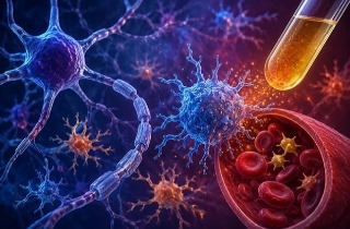
Further experiments demonstrated that ORF3a was being transported inside exosomes, microscopic membrane-bound particles released by infected cells. When researchers blocked exosome production, the amount of ORF3a reaching the liver dropped significantly. Fat accumulation, immune cell infiltration, and liver damage were also substantially reduced.
Exosomes Act Like Biological Delivery Vehicles
Exosomes are naturally produced by cells and serve as communication vehicles between organs. SARS-CoV-2 appears to exploit this system.
The researchers showed that ORF3a-loaded exosomes released fr
om lung tissue could travel through the bloodstream and become concentrated in the liver. Since the liver normally removes more than 90 percent of circulating exosomes from the blood, it becomes a prime target for these viral protein packages.
Once inside the liver, ORF3a interferes with the cell’s waste disposal and recycling systems. This prevents the normal breakdown of fat droplets, causing fat to accumulate within liver cells.
Over time, this excess fat triggers inflammation and activates cells involved in fibrosis, creating conditions that resemble early nonalcoholic fatty liver disease.
Human Liver Models Confirm the Findings
To determine whether the same process could occur in humans, scientists developed sophisticated laboratory-grown human liver organoids containing hepatocytes, macrophages, and hepatic stellate cells.
When ORF3a-containing exosomes were introduced into these miniature liver systems, the results closely mirrored those observed in animals.
Liver cells accumulated significantly more fat and triglycerides. Genes involved in fat production became highly active. Immune cells released elevated levels of inflammatory molecules including IL-1β, IL-6, and TNFα. At the same time, hepatic stellate cells transformed into scar-forming cells associated with fibrosis.
Remarkably, the researchers found that the ORF3a protein from SARS-CoV-2 was much more effective at causing these harmful changes than the similar ORF3a protein from the original SARS virus that caused the 2003 outbreak.
This Medical News report highlights how a single viral protein can influence organs far from the original site of infection without requiring the virus itself to spread throughout the body.
Potential Implications for Long COVID and Liver Health
The findings suggest that many cases of liver dysfunction seen during and after COVID-19 may not be caused by direct viral infection of the liver. Instead, they may result from viral proteins traveling through the body's communication networks.
Researchers believe that therapies designed to block ORF3a activity, prevent exosome-mediated transport, or interfere with the protein's interaction with host cellular machinery could help reduce COVID-19-related liver complications.
Conclusion
This study provides compelling evidence that mild COVID-19 can affect the liver through a previously underappreciated lung-liver communication pathway. The viral protein ORF3a was shown to travel from infected lungs to the liver inside exosomes, where it disrupted fat metabolism, promoted inflammation, and initiated fibrotic changes. These findings help explain why liver abnormalities are frequently observed even when the virus remains largely confined to the respiratory tract. The discovery also opens new opportunities for developing treatments aimed at blocking viral protein transport and protecting vulnerable organs from long-term COVID-19 complications.
The researchers were from the University of Southern California Keck School of Medicine (USA), Shanghai Children's Hospital and Shanghai Jiao Tong University School of Medicine (China), Shanghai Institute of Immunology (China), Cleveland Clinic Foundation (USA), Ruijin Hospital Shanghai Jiao Tong University School of Medicine (China), Tulane University School of Medicine (USA), the Shanghai Key Laboratory of Emergency Prevention, Diagnosis and Treatment of Respiratory Infectious Diseases (China), and the National Research Center for Translational Medicine at Shanghai (China).
The study findings were published in the peer reviewed journal: Cell Discovery.
https://link.springer.com/article/10.1038/s41421-026-00901-9
For the latest COVID-19 news, keep on logging to Thailand Medical News.
Read Also:
https://www.thailandmedical.news/articles/coronavirus
https://www.thailandmedical.news/articles/long-covid