Nikhil Prasad Fact checked by:Thailand Medical News Team Jun 02, 2026 1 month, 2 weeks, 6 days, 7 hours, 50 minutes ago
Medical News:
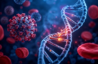
COVID-19 Recovery May Leave Hidden Brain Changes for Months
A new brain imaging study has found that COVID-19 may leave behind subtle but potentially important changes in the brain long after the infection has cleared. Researchers discovered evidence of altered brain communication networks and white matter abnormalities in people who had recovered from COVID-19, with the most pronounced effects seen in those who had experienced more severe illness requiring hospitalization.
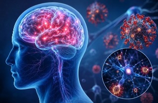
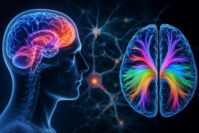 Advanced MRI scans reveal persistent brain network and white matter changes in people recovering from COVID-19,
especially after severe illness
The research was conducted by scientists from the Department of Electrical Engineering at the Indian Institute of Technology Delhi, India; the FMRIB Centre, Nuffield Department of Clinical Neurosciences at the University of Oxford, United Kingdom; and the Department of Biomedical Engineering at the New Jersey Institute of Technology (NJIT), United States.
Looking Beyond Recovery
Although most people recover from COVID-19, many continue to report lingering symptoms such as fatigue, memory problems, poor concentration, sleep disturbances, anxiety, and mood changes. These persistent issues, often referred to as Long COVID or Post-COVID Syndrome, have raised concerns that the virus may leave lasting effects on the brain.
To investigate this possibility, researchers used multiple advanced MRI techniques to examine the brains of 76 recovered COVID-19 patients and 51 healthy individuals who had never been infected. The study explored brain structure, white matter integrity, and functional communication between key brain regions.
Severe COVID Linked to Shrinkage in Key Brain Regions
One of the most notable findings involved the orbitofrontal cortex, a region located just above the eyes that plays an important role in decision-making, emotional regulation, reward processing, and social behavior.
When all recovered patients were analyzed together, researchers found no major differences in overall brain volume compared to healthy controls. However, after separating patients according to disease severity, a different picture emerged. Individuals who had been hospitalized during their COVID-19 illness showed significant volume loss in the orbitofrontal cortex and frontal pole compared to both healthy individuals and those who experienced milder infections.
These findings suggest that severe COVID-19 may accelerate damage or loss of brain tissue in regions involved in emotional and cognitive functioning.
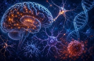
White Matter Damage Appears Widespread
The study revealed even stronger evidence of persistent abnormalities in the brain's white matter—the network of nerve fibers that connects different brain regions and allows them to communicate efficiently.
Researchers found widespread alterations in several major white matter pathways, including the inferior fronto-occipital fasciculus, corpus callosum, cingulum, internal capsule, and superior longitudinal fasciculus. These pathways a
re crucial for attention, memory, executive function, emotional processing, and sensory integration.
Many of these changes were detected through measurements known as fractional anisotropy and mean diffusivity, which provide clues about the health and organization of nerve fibers. The findings suggest that COVID-19 recovery may be associated with long-lasting microstructural disruptions within the brain's communication highways.
Interestingly, some regions showed increased connectivity-related measures while others showed reductions, indicating that the brain may be attempting to reorganize itself following injury or inflammation. The researchers believe some of these changes may reflect compensatory adaptations rather than straightforward damage.
Thalamus Shows Signs of Network Rewiring
Perhaps the most intriguing discovery involved the thalamus, a deep brain structure that acts as a central relay station, helping coordinate sensory, cognitive, and emotional information throughout the brain.
While researchers did not observe significant thalamic volume loss, they found clear evidence that communication between the mediodorsal thalamic nucleus and several cortical regions had changed after COVID-19 recovery.
Hospitalized patients demonstrated increased connectivity between the mediodorsal thalamus and areas involved in attention, visual processing, sensory integration, and higher-order cognition. Enhanced connections were observed with the anterior cingulate cortex, superior parietal regions, angular gyrus, occipital cortex, visual association areas, and other brain regions involved in processing information from the environment.
This
Medical News report notes that these findings are particularly important because the mediodorsal thalamus plays a major role in attention, memory, motivation, emotional regulation, and executive function—areas commonly affected in Long COVID patients.
Possible Explanation for Long COVID Symptoms
The researchers noted that fatigue was the most common lingering symptom among participants, affecting 68 percent of those reporting post-COVID symptoms. Other common complaints included poor sleep, reduced attention, headaches, muscle aches, and joint pain.
The observed abnormalities in fronto-limbic and thalamocortical networks may help explain why many individuals continue to experience cognitive and emotional difficulties months after apparent recovery.
Conclusions
The study provides compelling evidence that COVID-19 recovery does not always mean complete neurological recovery. While large-scale brain tissue loss was not seen in all recovered patients, significant changes were detected in critical brain communication networks, particularly among individuals who suffered more severe infections. The combination of orbitofrontal volume loss, widespread white matter abnormalities, and altered thalamic connectivity suggests that COVID-19 may trigger long-lasting reorganization of brain circuits involved in cognition, attention, memory, and emotional regulation. These findings support growing concerns that the neurological effects of COVID-19 can persist well beyond the acute illness and may contribute directly to the symptoms experienced by many Long COVID sufferers. Further long-term studies will be needed to determine whether these brain changes eventually resolve or become permanent.
The study findings were published on a preprint server and are currently being peer reviewed.
https://www.medrxiv.org/content/10.64898/2026.05.19.26353613v1
For the latest on Long COVID, keep on logging to Thailand
Medical News.
Read Also:
https://www.thailandmedical.news/articles/long-covid
https://www.thailandmedical.news/articles/coronavirus


 Share
Share
 Tweet
Tweet
 Share
Share