Nikhil Prasad Fact checked by:Thailand Medical News Team Jun 26, 2026 1 day, 3 hours, 51 minutes ago
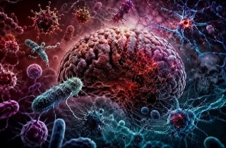
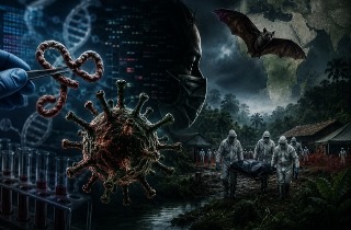
Medical News: As chikungunya virus continues to spread across the globe and causes increasingly severe neurological complications in some patients, scientists have uncovered a surprising difference in how two of the brain's most important cell types respond to the infection. The discovery could help explain why the virus damages the brain and may also open the door to new treatment strategies.
 Scientists discover that chikungunya virus activates completely different defense mechanisms in astrocytes
and neurons, revealing new clues to brain damage during infection
Researchers from the W. Harry Feinstone Department of Molecular Microbiology and Immunology, the Department of Biochemistry and Molecular Biology, the Department of Molecular Biology and Genetics, the McKusick-Nathans Department of Genetic Medicine, and the Department of Oncology at Johns Hopkins University, along with the Department of Physiology and Pharmacology at Oregon Health & Science University, found that astrocytes and neurons react to chikungunya virus in completely different ways despite both becoming infected.
The Brain's Cells Do Not Fight the Virus Equally
Chikungunya virus is best known for causing fever, rash, severe joint pain and long-lasting arthritis. However, mounting evidence shows that it can also invade the brain, leading to encephalitis, meningitis, cognitive decline and other neurological disorders. Understanding how the virus behaves inside brain cells has therefore become increasingly important.
The researchers compared astrocytes, which help protect and support nerve cells, with neurons, the specialized cells responsible for transmitting signals throughout the brain and nervous system.
Their experiments showed that astrocytes rapidly activated powerful antiviral defenses after infection. They switched on immune sensors including RIG-I and MDA5, activated IRF3, produced large amounts of type I interferons, and stimulated numerous interferon-responsive genes. These interferons act as natural warning molecules that alert neighboring cells and help slow viral spread.
Neurons, however, displayed a remarkably weak immune response. They failed to produce detectable type I interferons and showed little activation of many of the antiviral genes that became highly active in astrocytes.
A Completely Different Pathway Becomes Active
Instead of launching a strong immune response, infected neurons activated an entirely different process involving the enzyme PARP1.
The investigators discovered that chikungunya infection caused activation of PARP1 through caspase-3-driven cell death pathways. This led to widespread ADP-ribosylation, a chemical modification that changes the activity of many proteins inside cells.
Interestingly, this occurred without increasing production of most PARP genes themselves. In contrast, astrocytes increased expression of several interferon-stimulated PARP genes but showed very little overall ADP-ribosylation.
This striking contrast suggests that identical viral infections can trigger fundamentally different molecular prog
rams depending on the type of brain cell involved.
Viral Protein Helps Suppress Immune Defenses
The researchers also examined mutations within the virus's non-structural protein 3 macrodomain, a region already known to contribute to the virus's ability to cause neurological disease.
Viruses carrying weakened macrodomain mutations triggered significantly higher production of interferon, particularly IFN-beta, in astrocytes while simultaneously reducing viral replication. These findings suggest that the viral protein normally helps suppress the body's natural antiviral defenses, allowing the virus to reproduce more efficiently.
This
Medical News report highlights how the virus appears to manipulate different brain cells using distinct biological strategies, making infection far more complex than previously appreciated.
Important Clues for Future Treatments
The team also tested several PARP inhibitors. Blocking PARP activity produced opposite effects in different brain cells. In astrocytes, inhibiting PARP proteins allowed greater viral replication, indicating these enzymes normally help restrict infection. In neurons, however, PARP1 inhibition reduced viral replication, suggesting the virus exploits PARP1 activity inside nerve cells to support its own survival.
These opposite responses demonstrate why therapies targeting a single pathway may produce very different results depending on which brain cells are affected during infection.
Conclusion
The study provides compelling evidence that chikungunya virus does not interact uniformly with the brain. Instead, astrocytes mount an effective interferon-based antiviral response, while neurons activate PARP1-dependent pathways linked to cell injury and viral persistence. These discoveries significantly improve understanding of chikungunya-associated neurological disease and identify several promising molecular targets that could eventually lead to therapies capable of protecting the brain while limiting viral replication during severe infections.
The study findings were published in the peer reviewed journal: Microbiology Spectrum.
https://journals.asm.org/doi/10.1128/spectrum.04178-25
For the latest research on the Chikungunya Virus, keep on logging to Thailand
Medical News.
Read Also:
https://www.thailandmedical.news/news/chikungunya-virus-triggers-chronic-arthritis
https://www.thailandmedical.news/news/new-viral-blocking-compound-derived-from-the-phytochemical-coumarin-shows-promise-against-chikungunya-virus
https://www.thailandmedical.news/news/spirulina-algae-shows-powerful-effects-against-chikungunya-virus


 Share
Share
 Tweet
Tweet
 Share
Share