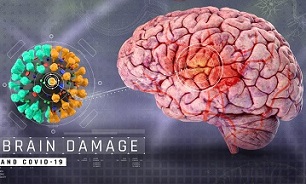
Great News! South Korean Study Shows That SARS-CoV-2 Is Literally Killing The Microglia Cells In The Host Brain! Hence The Various Arising Neurological Issues!
Source: Jan 11, 2022 4 years, 1 month, 1 week, 5 days, 14 hours, 37 minutes ago
More great news especially since the world is already full of stupid and ignorant lower life forms ie the pro-vaxxers, the anti-vaxxers, those that believe in ivermectin and fluvoxamine, those that believe in Fauci, those that believe and follow the so called ‘experts’ on twitter, those that think the SARS-CoV-2 coronavirus does not exists, those that think the pandemic will resolve to become an endemic, those that think asymptomatic and mild SARS-Cov-2 coronavirus infections are ok, those that believe in the Western mainstream and medical media, those that believe that wearing a N95 mask alone is going to protect you from the Omicron, those that believe in doctors alone without doing their own due diligence, those that believe in the WHO or US CDC, humans turning against humans, political leaders wanting to punish the unvaccinated, dictators imposing mandates and restrictions on fellow humans, etc and so on.
It is great to know that the SARS-CoV-2 coronavirus and various emerging variants are one way or another going to help deplete the human population especially with the long-term health complications or what we call as
long COVID. We are only just 24 months into the COVID-19 pandemic and already we are beginning to start seeing some of the more serious effects of long COVID-19 such as heart failures, kidney failures, liver disease, gastrointestinal issues and sepsis, strokes, colon cancer liver cancer, cerebral venous sinus thrombosis (CVST) etc. Most of which fortunately will end up in fatal outcomes but which the medical community and the health authorities are not linking to COVID-19!

Omicron is really god sent, as its sweeping fast in populations extremely fast and no one will be able to escape it.
Now reverting to the new but great study findings, researchers from the Korea Research Institute of Chemical Technology-South Korea, the College of Medicine at Yonsei University-South Korea and Arontier Co., Ltd-South Korea have in a new study found that the SARS-CoV-2 coronavirus is actually killing the microglia cells in the human host brain! This is also a major contributing factor as to why we are seeing a multitude of neurological and neuropsychiatric issues emerging from COVID-19 infections and also in Long COVID or PASC or Post-Acute Sequelae of SARS CoV-2 infection.
Microglia are a type of neuroglia (glial cell) located throughout the brain and spinal cord. Microglia account for 10–15% of all cells found within the brain. As the resident macrophage cells, they act as the first and main form of active immune defense in the central nervous system (CNS) and are actually the immune cells of the brain. Microglia (and other neuroglia including astrocytes) are distributed in large non-overlapping regions throughout the CNS. Microglia are key cells in overall brain maintenance ie they are constantly scavenging the CNS for plaques, damaged or unnecessary neurons and synapses, and infectious agents. Since these processes must be efficient to prevent potentially fatal damage, microglia are extremely sensitive to even small pathological changes in the CNS
To date, numerous evidence suggests that SARS-CoV-2 infection causes various neurological symptoms in COVID-19 patients. The most dominant immune cells in the brain are microglia. Yet, the relationship between neurological manifestations, neuroinflammation, and host immune res
ponse of microglia to SARS-CoV-2 has not been well characterized.
The study findings of this new research shockingly reveal that SARS-CoV-2 can directly infect human microglia, eliciting M1-like pro-inflammatory responses, followed by cytopathic effects.
Most importantly, SARS-CoV-2 infected human microglial clone 3 (HMC3), leading to inflammatory activation and cell death. RNA-seq analysis also revealed that ER stress and immune responses were induced in the early and apoptotic processes in the late phase of viral infection. SARS-CoV-2-infected HMC3 showed the M1 phenotype and produced pro-inflammatory cytokines such as interleukin (IL)-1β, IL-6, and tumour necrosis factor α (TNF-α), but not the anti-inflammatory cytokine IL-10.
It was found that after this pro-inflammatory activation, SARS-CoV-2 infection promoted both intrinsic and extrinsic death receptor-mediated apoptosis in HMC3.
The study team using K18-hACE2 transgenic mice found that murine microglia were also infected by intranasal inoculation of SARS-CoV-2. This infection induced the acute production of pro-inflammatory microglial IL-6 and TNF-α and provoked a chronic loss of microglia.
The study findings suggest that microglia are potential mediators of SARS-CoV-2-induced neurological problems and, consequently, can be targets of therapeutic strategies against neurological diseases in COVID-19 patients.
It should also be noted that recent studies reported neurological manifestations and complications in COVID-19 patients, which are associated with neuroinflammation. As microglia are the dominant immune cells in brains, it needs to be elucidate the relationship between neuroinflammation and host immune response of microglia to SARS-CoV-2.
The study findings already showed that SARS-CoV-2 can directly infect human microglia with cytopathic effect (CPE) using human microglial clone 3 (HMC3). The infected microglia were promoted to pro-inflammatory activation following apoptotic cell death. This pro-inflammatory activation was accompanied by the high production of pro-inflammatory cytokines, and led to neurotoxic-M1 phenotype polarization.
Furthermore, the HMC3 cells are susceptible to SARS-CoV-2 and exhibit the CPE, which can be further used to investigate cellular and molecular mechanisms of neuroinflammation reported in COVID-19 patients.
The study findings were published on a preprint server and are being peer reviewed for publication into a leading scientific journal.
https://www.biorxiv.org/content/10.1101/2022.01.04.475015v1
Microglia are macrophage-like immune cells in the brain and central nervous system (CNS) that maintain brain homeostasis and are known to act in response to injury and inflammation rapidly.
Typically, in response to immunological stimulus, microglial cells adopt an amoeboid morphology and release interleukins (IL) like IL-1β and IL-6 and tumor necrosis factor–α (TNFα).
Importantly microglia exhibit dual phenotypes when activated. Whereas M1 is considered the classically activated state, M2 is the alternately activated state.
It is known that the M1 phenotype is involved in neuroinflammation and is neurotoxic; conversely, the M2 phenotype is neuroprotective.
Though much is known about microglial activation and response, more research is required to characterize and understand the microglial host-immune response in patients infected with SARS-CoV-2.
The study team investigated the factors driving neuroinflammation and other neurological complications in COVID-19 patients in the present study.
This research was primarily conducted in response to several reports of microgliosis, accumulation of immune cells, and microglial nodules in the medulla oblongata and cerebellar dentate nuclei in the brains of deceased COVID-19 patients that arise due to massive activation of microglial cells.
The study team investigated whether SARS-CoV-2 can infect human microglial cells by inoculating the human embryonic primary microglia (HMC3) with one multiplicity of infection (MOI) of SARS-CoV-2. In addition, other SARS-CoV-2-susceptible cell lines like Caco-2 and Vero E6 cells were also infected.
The study team found that SARS-CoV-2 infected HMC3 and that this infection triggered the death of HMC3 cells and exhibited a cytopathic effect (CPE). The authors also investigated whether SARS-CoV-2 infection elicited an M1 or M2 phenotype of microglia and analyzed the differentially expressed genes (DEGs) associated with microglial polarization.
It was found that an increase in ribonucleic acid (RNA), expression levels of M1 phenotype-related genes like IL-1β, IL-6, and CXCl1 was observed.
This suggests that SARS-CoV-2 infection induces a pro-inflammatory activation leading to the M1 phenotype in HMC3.
The study team further determined the mechanism of death of HMC3 cells triggered due to SARS-CoV-2 infection.
Western blot analysis revealed that apoptotic proteins associated with both intrinsic and extrinsic pathways of apoptosis were elicited in the SARS-CoV-2-infected HMC3 cells.
Interestingly death receptor (DR)-mediated proteins of the extrinsic apoptotic pathway such as Fas, death receptor 4 (DR4), DR5, and TNF receptor 2 (TNFR2) were observed on the Western blots.
Furthermore, the expression of Bcl-2 (apoptosis suppressor) decreased while that of Bim, Bid, and Bax increased.
These study findings reveal that SARS-CoV-2 induces cell death of HMC3 cells through both pathways of apoptosis.
It should be noted that several other RNA viruses such as the Zika virus (ZIKV) and vesicular stomatitis virus (VSV) cause pyroptosis, or inflammatory cell death, in most immune cells.
The study team ascertained whether pyroptosis is induced by SARS-CoV-2 infection in HMC3 and reported that pyroptosis due to SARS-CoV-2 was not detected in HMC3 cells and concluded that cell death was due to apoptosis.
The study team subsequently had transgenic mice (K18-hACE2) expressing human ACE2 with a cytokeratin-18 gene promoter, infected with 2 x 10⁴ plaque-forming units (PFUs) of SARS-CoV-2. After six days of infection, weight loss of about 20% of their body weight was observed in infected mice, and viral RNA was detected in their brains.
The study findings reveal that SARS-CoV-2 infects microglia and induces its subsequent activation and transformation into a pro-inflammatory M1 phenotype.
In addition, it was noted that microglial cell death due to SARS-CoV-2 infection is apoptotic, and both extrinsic and intrinsic ways of apoptosis were observed.
Importantly an increase in neurotoxic microglia (M1 cells) can lead to other neurological complications, such as the activation of astrocytes and T-lymphocytes, which can cause neuronal damage and death. Furthermore, the blood-brain barrier could be disturbed due to the release of cytokines by microglia that might cause additional neurological symptoms in COVID-19 patients.
Further in vivo investigations in transgenic mice reported the infection of microglia by SARS-CoV-2, which induced the release of pro-inflammatory cytokines and subsequently caused chronic loss of microglia.
All these study findings suggest a lack of immune response in the brain; therefore, the increased viral replication may lead to different neurological manifestations.
Importantly the observations made in this study suggest that microglial cells could be targeted for therapeutic interventions in COVID-19 patients presenting with neurological symptoms.
With new studies showing that viral persistence can last for months even after being deemed as recovered by stupid and inaccurate nasal or mouth swab tests, we can expect to see more people suffering a variety of neurological and neuropsychiatric conditions in coming months and years and hopefully an increase in fatal strokes etc.
https://www.thailandmedical.news/news/breaking-u-s-nih-study-shockingly-reveals-sars-cov-2-viral-persistence-throughout-human-body-and-in-the-brain-even-in-those-who-were-asymptomatic
The American army, DARPA, Fauci and the Democrats should be praised for developing such as effective bioweapon as the SARS-Cov-2 virus, its just unfortunate that they lost control of it but for eugenics and those in favor of genocides like a famous tech billionaire, SARS-CoV-2 is his dream come true.
For those wanting to know how to survive the ongoing and coming COVID-19 surges and prevent the various long COVID-19 afflictions, join our private community where we share the latest prophylactics, therapeutics and various protocols in private. Entry is via paid membership only and cost US$2,000. Those interested drop us an email.
For more great news on the SARS-CoV-2 and eradication of the stupid and ignorant, keep on logging to Thailand Medical News.
