Nikhil Prasad Fact checked by:Thailand Medical News Team Oct 04, 2025 9 months, 1 week, 2 days, 21 hours, 3 minutes ago
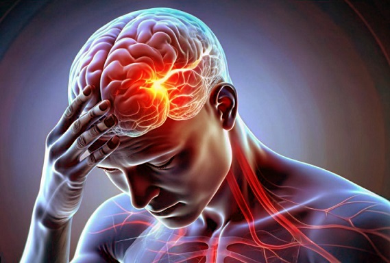
Medical News: A closer look at how COVID 19 harms brain health
Scientists from the University of Illinois Chicago, Department of Anatomy and Cell Biology and the Department of Microbiology and Immunology at the same institution have uncovered new evidence that SARS-CoV-2, the virus behind COVID-19, directly disrupts crucial signaling in brain cells known as astrocytes. These cells are vital for maintaining the blood–brain barrier, reducing inflammation, and supporting normal brain function. This
Medical News report reveals that the findings may help explain why so many people develop brain fog, memory issues, and other neurological symptoms during COVID-19 and Long COVID.
 SARS-CoV-2 Disrupts Brain Cell Pathways Fueling Long-Term Neurological Problems
The study and what researchers found
SARS-CoV-2 Disrupts Brain Cell Pathways Fueling Long-Term Neurological Problems
The study and what researchers found
The team used laboratory-grown human astrocytes derived from stem cells to test whether the virus could infect them. They confirmed that SARS-CoV-2 not only infected the cells but also continued releasing viral particles for up to 96 hours. This sustained infection suggests astrocytes could play a hidden role in lingering neurological damage.
The researchers then focused on the Wnt/β-catenin pathway, a molecular system that helps regulate the blood–brain barrier and supports neuron growth and repair. Their experiments revealed several alarming changes:
-Levels of β-catenin, a key protein in this pathway, dropped by about 50 percent after infection.
-Wnt3a and Wnt10b, molecules important for brain cell survival and repair, were also reduced by 70 to 75 percent.
-In contrast, Wnt7b was boosted, which may represent the brain’s attempt to repair blood–brain barrier damage.
-Another major change was the activation of CXCL10, a chemical messenger linked to inflammation, which rose four-fold in infected astrocytes.
Interestingly, despite these strong shifts in cellular function, the virus did not appear to kill the astrocytes directly. Instead, it altered how they behaved, making them more inflammatory and less protective.
Why these findings matter
Astrocytes normally act as guardians of the brain, releasing signals that protect blood vessels and keep harmful substances from leaking into brain tissue. When SARS-CoV-2 disrupts these pathways, the blood–brain barrier may weaken, allowing inflammation to spread. The drop in Wnt3a and Wnt10b may further reduce the brain’s ability to repair itself after infection. At the same time, higher levels of CXCL10 suggest a state of chronic inflammation, which is known to play a role in conditions like Alzheimer’s, Parkinson’s, and multiple sclerosis.
These results strengthen the idea that Long COVID brain symptoms are not just psychological but rooted i
n real biological damage. The findings also highlight the Wnt/β-catenin pathway as a possible target for future treatments designed to reduce neuroinflammation and protect brain health in COVID-19 survivors.
Conclusion
This important research shows that SARS-CoV-2 does not simply infect the lungs but can infiltrate brain cells and interfere with delicate molecular systems. By reducing protective proteins and boosting inflammatory signals, the virus may set the stage for long-lasting neurological complications. The study helps explain why so many patients report brain fog and memory issues months after infection. Protecting or restoring the Wnt/β-catenin pathway could become a new approach in managing Long COVID and other virus-linked brain disorders.
The study findings were published in the peer reviewed journal: Pathogens.
https://www.mdpi.com/2076-0817/14/10/994
For the latest COVID-19 News, keep on logging to Thailand
Medical News.
Read Also:
https://www.thailandmedical.news/news/enlarged-choroid-plexus-in-long-covid-linked-to-cognitive-and-brain-changes
https://www.thailandmedical.news/news/new-study-validates-that-sars-cov-2-infects-brain-neurons-through-cathepsins-and-triggers-alzheimer-like-changes
https://www.thailandmedical.news/news/covid-19-infection-tied-to-higher-risk-of-vascular-dementia-in-older-adults


 Share
Share
 Tweet
Tweet
 Share
Share


