BREAKING! U.S Department Of Defense Study Finds That COVID-19 Infected Individuals Younger Than 60 Years Are Developing Beta-Amyloid Deposits!
Source: Beta-Amyloid-COVID-19 Jan 26, 2022 4 years, 4 months, 3 weeks, 5 days, 3 hours, 51 minutes ago
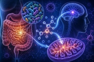
Beta-Amyloid-COVID-19: A new study led by the U.S. Department of Defense involving researchers, famous neurodegenerative disease specialists and pathologists from the Uniformed Services University of the Health Sciences- Maryland, the Icahn School Of medicine At Mount Sinai-New York and Columbia University-New York have in a new study alarmingly found that COVID-19 infected individuals younger than 60 years are developing beta-amyloid deposits!
In the study started with a 58-year-old woman who died of COVID infection was included as a control subject in an ongoing autopsy study of the neuropathology of military traumatic brain injury (TBI).
Surprisingly an immunohistochemical stain for β-amyloid done only because that stain is performed on all the cases in the TBI study resulted in the serendipitous observation of numerous focal β-amyloid deposits in the neocortex of this relatively young woman with no significant neurologic history.
The immunohistochemical study was replicated in a second laboratory using a different anti-amyloid monoclonal antibody with the same result. The deposits failed to stain with thioflavin-S.
The
Beta-Amyloid-COVID-19 study team subsequently found identical findings in 10 out of 10 additional autopsies of severely ill, hospitalized COVID patients younger than 60 years.
All these patients were all severely ill COVID patients who died of their disease and they are probably not representative of the typical COVID patient who often has a relatively mild clinical course and no need for hospitalization.
However, one COVID patient in the ongoing autopsy study of the neuropathology of military TBI was a 39-year-old-man was self-isolating at home when he became acutely short of breath and died the same day in spite of advanced cardiac life support efforts.
The study team interpreted this case as an example of a typical mild COVID infection which was probably complicated by a COVID-associated pulmonary embolus. At autopsy this patient had only extremely rare focal β-amyloid deposits. The focal β-amyloid deposits described here are not unique to COVID patients.
Further examination of other patients who died in the pre-COVID era with adult respiratory distress syndrome also revealed these amyloid deposits, suggesting a link to hypoxia.
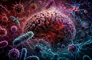
COVID encephalopathy and/or the persistent cognitive impairment in COVID survivors is well documented and becoming a serious public health concern.
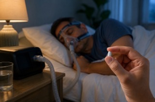
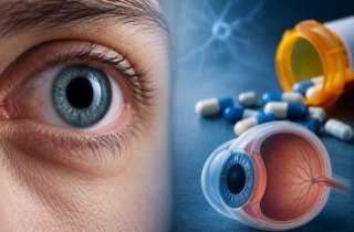
The study team speculates that the β-amyloid deposits described here may play a role in the pathophysiology of that syndrome. If that is true, COVID encephalopathy may respond to treatment with anti-amyloid therapeutics.
The study findings were published on a preprint server by lancet and is currently being peer reviewed.
https://papers.ssrn.com/sol3/papers.cfm?abstract_id=4003213
The debut of the SARS-CoV-2 coronavirus in Wuhan-China in December 2019 and subsequent spread of the virus
globally by the irresponsible and selfish Chinese people who were serving like human vectors of the disease led to the current global COVID-19 pandemic.
Aside from the acute effects of COVID-19, which can be severe and even fatal in a significant minority of cases, long-term effects of COVID-19 can occur, even after the patient has recovered.
It is now being projected from an initial 33 percent, that up to 87 percent of all infected with the SARS-CoV-2 coronavirus, irrespective if they were asymptomatic, or had mild, moderate or even severe symptoms or conditions would ultimately develop one of more health issues associated with Long COVID.
Long COVID often manifests itself in protean ways. One such symptom is brain fog, or cognitive dysfunction, which is attributed to neurologic damage directly or indirectly caused by the infection. Recovered COVID-19 patients are also at risk of several neuropsychiatric conditions, including dementia, probably due to a variety of underlying pathogenetic mechanisms.
This new study findings by researchers associated with the U.S. Department of Defense describes the unexpected but valuable finding that patients who died as a result of COVID-19 exhibit large amounts of focal β-amyloid deposits in their neocortical tissues upon autopsy.
The data correlation with the findings from the brains of non-COVID-19 patients who experienced markedly hypoxic conditions indicates that the amyloid deposition is not a specific sign of SARS-CoV-2 infection, but rather could be a response to brain hypoxia.
The study team autopsied COVID-19 patients and examined their neocortex for brain deposits of beta-amyloid. The first patient was a 58-year-old female with a history of psychiatric disorders who died of COVID-19 soon after her diagnosis. Following the autopsy, the diagnosis of intoxication with methadone, gabapentin, and hydroxyzine was made, along with the effects of COVID-19.
However, despite the presence of psychiatric history, including depression, suicidal ideation, and post-traumatic stress disorder (PTSD), this patient did not show any neurological abnormalities and had a normal cognitive function. At some point before her death, she had suffered a traumatic brain injury that was not associated with any neurologic sequelae.
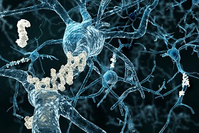
The detailed brain autopsy showed no gross abnormalities; however, numerous deposits of β-amyloid were found scattered in the neocortex and as puncta within cell bodies. Fewer, but still significant, flat plaques or deposits of the protein were present in the hippocampus, with a small number of cells containing the tau protein in one region.
Interestingly, tiny foci of the tau protein were also found in a few neurites in the same region. Many fibers in the occipital white matter and entorhinal cortex were also positive for tau immunostaining.
Considering the absence of neurofibrillary tangles or other evidence of neurodegenerative conditions, the presence of these deposits was remarkable.
Furthermore normal-appearing microglia were also stained. However, unlike Alzheimer’s disease (AD) patients, the β-amyloid deposits were not stained with thioflavin-S, which is why they have not included plaques in the final description. Furthermore, these deposits were not bound by the monoclonal antibody 22C11.
Importantly the lack of 22C11 indicates the absence of the amino acid residues at position 66-81 in the N-terminus of the amyloid precursor protein. In contrast, the stain was taken up, though weakly, by the puncta within the cells.
As a result of these findings, the content of the deposits appears to consist of a peptide that has been broken down by proteolysis.
Hence, these deposits could be the precursor of the β-amyloid plaques or of plaques made up of a similar molecule that was identified prior to its polymerization, subsequent to which the beta-pleated sheet structure would develop.
In order to evaluate for the presence of β-amyloid in other cases of fatal COVID-19, the study team examined another ten patients under the age of 60 years.
Shockingly the same findings were identified in all ten, with one individual also showing rare neuropil threads positive for the tau protein. Though small infarcts were found in several patients, none of these lesions were found in the stained sections.
The study team also examined the brain tissue of five patients who had died of Adult Respiratory Distress Syndrome (ARDS) in the pre-pandemic years. To this end, focal deposits of thioflavin-negative and β-amyloid immunoreactive protein were observed in the frontal cortex that could not be distinguished from those of the index COVID-19 patient.
The study team also looked at the biopsy results of several other patients who died of acute to subacute infarcts. Both showed similar amyloid deposits to the above two categories.
A detailed routine immunohistochemical assay had earlier been carried out on an unrelated cohort of patients with military traumatic brain injury (TBI), which was used as a control cohort.
Out of 61 individuals, all without severe respiratory disease, only five had this type of amyloid deposit, all between January 1, 2020, and December 31, 2020.
Importantly this indicates that young people rarely develop these amyloid deposits and that their presence is common in severe respiratory distress.
It should be noted that the clinical histories of these five patients had not been completely elucidated by the time of publication. However, this observation shows that young patients do not typically produce amyloid deposits and that the deposits that were found in COVID-19 and ARDS patients are not artefactual.
It was reported that during the late phase of this study, another COVID-19 patient died of rapidly progressing breathlessness on the fifth day after testing positive. The patient, a 39-year-old Marine, was obese but a full autopsy was not done. However, the brain was examined and the scientists observed microthrombi and microbleeds, both of which were observed in other fatal COVID-19 cases.
The β-amyloid plaques seen in the index patient were so rare in this case that they likely would have been missed without the careful search for them.
This patient’s death was attributed to a probable pulmonary embolus complicating a mild COVID-19 infection.
The study findings show the presence of β-amyloid deposits in young COVID-19 patients, as well as non-COVID-19 patients with hypoxia affecting the brain.
The correlation between these deposits and cognitive impairment, along with the neurologic deficits seen in some long COVID patients, appears to indicate that this is not a virus-specific lesion but a reflection of hypoxic brain damage.
The study finding supports this conclusion that all ten patients had some amount of hypoxia, which was seen in the brain as extensive hypoxic changes.
Possible explanations for the observation is that extensive hypoxic changes could lead to the release of hypoxia-inducible factor (HIF)-1α, which increases the production of β-amyloid. At the same, hypoxia reduces the rate at which this protein breaks down, thus increasing the level of the protein.
The study team says that further longitudinal prospective studies must be carried out to define the period over which these lesions survive and whether they are found in cases of mild infection and other convalescent patients. It will be important to determine how frequently these deposits occur in the brains of patients who recover from COVID-19 and then expire months or years later from other causes.
Yet another possible explanation is that these protein deposits herald the formation of amyloid plaque. The pathophysiology of neuropsychiatric symptoms in this viral infection may have been partly explained by these findings.
However, the study findings may also support the use of anti-amyloid in treating COVID-19 encephalopathy in the near future.
For more about
Beta-Amyloid-COVID-19, keep on logging to Thailand Medical News.


 Share
Share
 Tweet
Tweet
 Share
Share