Nikhil Prasad Fact checked by:Thailand Medical News Team Jul 20, 2026 1 month, 2 weeks, 1 day, 11 hours, 37 minutes ago
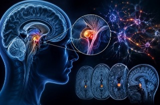
Medical News: Scientists Discover That Monitoring the Brain’s “Little Brain” Could Help Predict Neurological Diseases
The cerebellum, often referred to as the brain’s “little brain,” has traditionally been associated with balance and movement. However, a new scientific review suggests this small structure may hold valuable clues to the health of the entire brain. Researchers now believe that monitoring its electrical activity using Electrocerebellography (ECeG) could one day help doctors detect neurological disorders earlier and better understand how the brain functions in both health and disease.
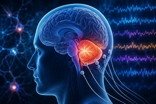 Researchers say Electrocerebellography (ECeG) could become a valuable tool for detecting early brain disorders by monitoring electrical activity in the cerebellum
The study was conducted by researchers from the Department of Neurophysiology, Institute for Biological Research “Siniša Stanković” – National Institute of the Republic of Serbia, University of Belgrade, Serbia. Their review examined research spanning the past three decades on cerebellar electrical activity and its relationship with numerous neurological and psychiatric conditions.
The Cerebellum Does Much More Than Control Movement
For many years, the cerebellum was thought to serve mainly as the brain's movement control center, helping coordinate posture, balance, and muscle activity. Scientists now know that it also plays major roles in memory, attention, speech, emotional regulation, social behavior, auditory processing, and learning.
Because the cerebellum has extensive connections with almost every major brain region and possesses remarkable synaptic plasticity—the ability to adapt and reorganize its neural connections—it offers a unique opportunity to observe changes occurring throughout the brain. Researchers believe alterations in its electrical signals may mirror widespread changes taking place long before obvious symptoms develop.
Electrical Brain Rhythms Could Become Powerful Biomarkers
Unlike conventional electroencephalography (EEG), which mainly records electrical activity from the surface of the cerebral cortex, Electrocerebellography (ECeG) specifically records electrical signals generated by the cerebellum. Although the technique has received relatively little attention over the years, growing evidence suggests it may become an important diagnostic and research tool.
Scientists found that the cerebellum produces distinct rhythmic electrical patterns depending on what the brain is doing. Slow theta rhythms appear to support sensory processing and learning, while beta and gamma rhythms help coordinate communication between different brain regions and control precise movement timing. Extremely fast oscillations are believed to synchronize Purkinje cells, allowing the brain to execute rapid, coordinated movements with remarkable precision.
Importantly, these rhythmic patterns change significantly during sleep, anesthesia, brain injury, epilepsy, and neurodegenerative diseases, making them potential biological markers for monitoring brain health.
r />
Study Highlights Expanding Clinical Potential
This Medical News report highlights how researchers increasingly view cerebellar electrical activity as a reflection of the functional connectivity between the cerebellum and the rest of the brain. Rather than acting independently, the cerebellum continuously exchanges information with multiple brain networks involved in cognition, emotion, and movement, meaning subtle electrical changes may provide early warning signs of disease.
Study Links ECeG Changes to Numerous Brain Disorders
The review describes compelling evidence connecting abnormal cerebellar electrical activity with many neurological and psychiatric disorders.
In epilepsy, patients often develop cerebellar degeneration, altered connectivity, and abnormal electrical rhythms. Although seizures usually begin in the cerebral cortex, the cerebellum appears to influence seizure activity and may even become a therapeutic target for future neurostimulation treatments.
Following traumatic brain injury, researchers observed long-lasting increases in gamma-frequency activity and reductions in spontaneous cerebellar electrical activity. These changes persisted long after the initial injury, suggesting ECeG could help monitor both acute damage and long-term recovery.
The findings are equally striking for Alzheimer's disease. Patients and experimental models consistently demonstrate increased slow-frequency brain activity together with reductions in faster oscillations. The review also describes loss of Purkinje cells, cerebellar atrophy, amyloid plaque accumulation, and declining communication with memory networks, indicating that cerebellar dysfunction develops alongside cognitive decline.
In Parkinson's disease, patients exhibit slower brain rhythms, reduced cerebellar theta activity during movement and thinking tasks, and abnormal high-frequency firing in deep cerebellar neurons. The researchers note that therapies such as deep brain stimulation and levodopa may partly work by restoring communication involving cerebellar circuits.
The review further summarizes evidence linking cerebellar electrical abnormalities with schizophrenia, depression, anxiety disorders, autism spectrum disorder, and disturbances in emotional regulation, demonstrating that the cerebellum contributes far more to mental health than previously recognized.
Sleep and Anesthesia Reveal Additional Insights
Sleep studies showed that cerebellar electrical activity changes throughout every stage of sleep. Activity decreases during deep non-REM sleep but becomes more active during REM sleep, when dreaming occurs. The cerebellum also communicates closely with the hippocampus and cerebral cortex during sleep, helping regulate memory consolidation and learning.
Researchers also found that common anesthetic agents consistently alter cerebellar electrical rhythms, producing dominant slow delta and theta oscillations while temporarily disrupting normal communication between cerebellar neurons. These findings may explain why anesthesia can temporarily impair coordination, cognition, and sleep quality after surgery.
Conclusions
The growing body of evidence strongly suggests that Electrocerebellography (ECeG) could evolve into an important biomarker for brain health. While additional human studies are still needed, the technology shows considerable promise for detecting early neurological changes, monitoring disease progression, evaluating treatment responses, and providing researchers with a deeper understanding of how brain networks function across both healthy and diseased states.
The study findings were published in the peer reviewed journal: Brain Sciences.
https://www.mdpi.com/2076-3425/16/7/758
For the latest on brain health issues, keep on logging to Thailand Medical News.
Read Also:
https://www.thailandmedical.news/articles/alzheimer,-dementia-




 Share
Share
 Tweet
Tweet
 Share
Share