BREAKING! SARS-CoV-2 Persists In Intestinal Enterocytes Up To 7 Months After Symptom Resolution. Viral Persistence Is Real And A Serious Issue!
Source: SARS-CoV-2 Viral Persistence Dec 25, 2021 4 years, 1 month, 4 weeks, 10 hours, 15 minutes ago
SARS-CoV-2 Viral Persistence: A study by researchers from Icahn School of Medicine at Mount Sinai -New York lead by Dr Minami Tokuyama has alarmingly found that the SARS-CoV-2 coronavirus persist in intestinal enterocytes up to 7 months after symptom resolution. The study which is still ongoing is finding that in some cases the viral persistence is even longer.
While you are here, for this Christmas, do something charitable and generous by making a donation to support this website and all our reserach and community initiatives. Thank You. https://www.thailandmedical.news/p/sponsorship

The preliminary study findings which have to be published as the study is ongoing, was released as an abstract in the Conference on Retroviruses and Opportunistic Infections (CROI) platform.
https://www.croiconference.org/abstract/sars-cov-2-persists-in-intestinal-enterocytes-up-to-7-months-after-symptom-resolution/
The host proteins ACE-2 and TMPRSS2 facilitate SARS-COV-2 infection and are expressed in the lungs as well as the intestinal tract, particularly in the small bowel.
Gastrointestinal symptoms represent the most common extrapulmonary manifestation of COVID-19. Viral RNA has been isolated from fecal samples from COVID-19 patients, where it can persist longer than detection in nasopharyngeal swabs. While SARS-CoV-2 infection of enterocytes has been demonstrated in vitro, in vivo studies are lacking.
The study involved small intestinal biopsies from patients who underwent clinically indicated endoscopic procedures after a positive SARS-COV-2 nasopharyngeal swab (n=27) or were found to have positive serology (n=2) were analyzed by immunofluorescence (IF) (n=25) and electron microscopy (EM) (n=14) for the presence of virus. Clinical details were also collected.
Sixteen of 29 patients had detectable SARS-CoV-2 antigen by either IF or EM.
The
SARS-CoV-2 Viral Persistence study findings showed that the virus was restricted to the epithelium and patchy in distribution. Virus was detected as soon as 15 days after symptom onset and persisted up to 6 months after symptom resolution. Five patients were nasopharyngeal swab positive at the time of procedure and, of these, 4 had detectable antigen on biopsy. Despite the presence of virus, only 9/16 patients had any signs of inflammation on histology, and when present, this was mild. In two patients where virus was present at 3 months and 4 months, additional biopsies were obtained at 7 months and 6 months, respectively.
Importantly viral antigen was persistently detected in both patients and both patients were nasopharyngeal swab negative for all procedures. Interestingly, only 37.5% (6 of 16) of patients with virus detected in the small bowel had GI symptoms (diarrhea, nausea or vomiting) during their acute COVID-19 illness as compared to 46.1%
(6/13) of patients where no virus could be detected in the intestines.
The study findings confirm that SARS-COV-2 infects enterocytes in humans in vivo and can persist in the intestines up to 7 months following symptoms resolution. This persistence is not associated with an overt inflammatory infiltrate and does not appear to correlate with presence of GI symptoms in the acute COVID-19 setting.
The team also appeals that more urgent detailed studies are conducted with regards to viral persistence in other parts of the human hosts.
The current method of using nasal swabs and lack of any existing symptoms as a means of determining COVID-19 recovery is totally inaccurate and misleading.
Viral persistence could be responsible for a variety of Long COVID-19 issues.
Despite other past studies supporting viral persistence in the intestinal tract, very little had been done to address this growing threat.
https://www.ncbi.nlm.nih.gov/pmc/articles/PMC7184976/
https://pubmed.ncbi.nlm.nih.gov/32969768/
https://www.immunology.ox.ac.uk/covid-19/literature-digest-old/sars-cov-2-may-persist-in-digestive-tract-longer-than-respiratory-tract
https://virologyj.biomedcentral.com/articles/10.1186/s12985-021-01599-9
https://www.frontiersin.org/articles/10.3389/fimmu.2021.674074/full
https://gut.bmj.com/content/69/6/973
https://www.frontiersin.org/articles/10.3389/fmed.2020.00562/full
https://academic.oup.com/cid/article/73/3/361/5868547
https://www.preprints.org/manuscript/202002.0354/v1
https://onlinelibrary.wiley.com/doi/pdf/10.1111/codi.15138
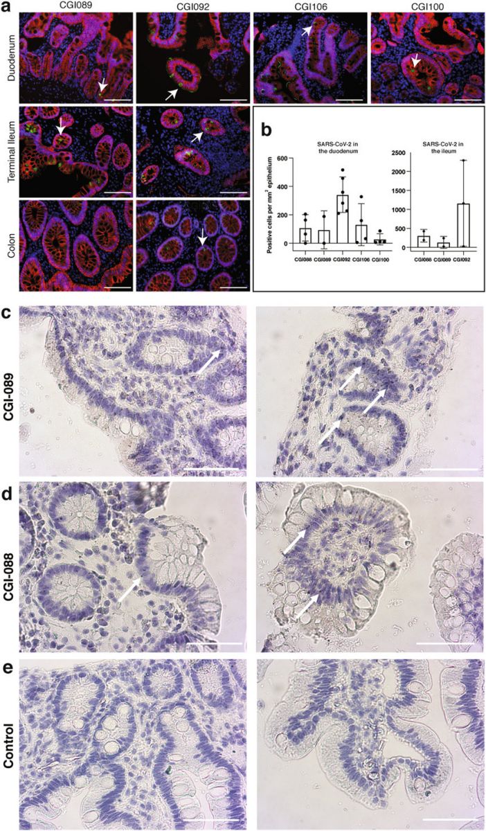
 SARS-CoV-2 antigen and RNA is detectable in different intestinal segments in several individuals convalescent from COVID-19 a, Immunofluorescence images of biopsy samples in the gastrointestinal tract in different individuals are shown. Staining is for EPCAM (red), DAPI (blue) and SARS-CoV-2 nucleocapsid (green). Samples are derived from intestinal biopsies from four participants (CGI-089, CGI-092, CGI-100 and CGI-106) taken at least three months after COVID-19 infection. Arrows indicate enterocytes with detectable SARS-CoV-2 antigen. Scale bars, 100 μm. The experiments were repeated independently at least twice with similar results. b, Quantification of SARS-CoV-2-positive cells by immunofluorescence. The number of cells staining positive for the N protein of SARS-CoV-2 per mm² of intestinal epithelium is shown. The graphs show biopsy samples from the indicated individuals of the duodenum (left) and terminal ileum (right). Black dots represent the number of available biopsy specimen for each individual from the respective intestinal segment (CGI-088, n = 4 duodenal and n = 2 ileal; CGI-089, n = 2 duodenal and n = 2 ileal; CGI-092, n = 6 duodenal and n = 3 ileal; CGI-106, n = 4; CGI-100, n = 4). Boxes represent median values and whiskers the 95% confidence interval. c, d, SARS-CoV-2 viral RNA was visualized in intestinal biopsies of participant CGI-089 (c) and CGI-088 (d) using in situ hybridization. SARS-CoV-2 genomic RNA (black) and haematoxylin and eosin staining by smFISH–immunohistochemistry technique in the duodenum (left) or terminal ileum (right). Arrows indicate enterocytes with detectable SARS-CoV-2 RNA. e, Pre-COVID-19 control individuals show no detectable SARS-CoV-2 viral RNA in duodenal (left) or ileal (right) biopsies. Scale bars, 100 μm. The experiments were repeated independently three times with similar results.
Thailand Medical News has also received reports that the WHO, the US CDC and US NIH despite acknowledging the seriousness of Long COVID or PASC (Post-Acute Sequelae of COVID-19) are deliberately trying to down play the seriousness of viral persistence and Long-COVID as it will badly reflect on the many wrong strategies that they gave undertaken since the beginning of the pandemic including the current diagnostics used and also the fact that they do not want the masses to panic. The excess deaths arising as a result SARS-CoV-2 persistence such as heart failures, acute kidney injuries, serious gastrointestinal issues leading to sepsis should all be carefully investigated as it is believed that the excess deaths rates as a result of SARS-CoV-2 persistence is really significant and far more than reported COVID-19 deaths!
While you are here, for this Christmas, do something charitable and generous by making a donation to support this website and all our research and community initiatives. Thank You. https://www.thailandmedical.news/p/sponsorship
For more about
SARS-CoV-2 Viral Persistence, keep on logging to Thailand Medical News.
Read Also:
https://www.thailandmedical.news/news/nice-neurologists-finds-replicable-sars-cov-2-rna-in-cerebrospinal-fluid-of-a-woman-114-days-after-she-was-diagnosed-with-asymptomatic-covid-19
https://www.thailandmedical.news/news/yet-another-alarming-research-finding-persistence-of-sars-cov-2-s1-protein-found-in-cd16-monocytes-of-post-covid-19-patients-up-to-15-months-after-so-
https://www.thailandmedical.news/news/children-s-hospital-los-angeles-led-study-alarmingly-shows-sars-cov-2-causing-viral-persistence-in-children-and-young-adults-with-cancer
https://www.thailandmedical.news/news/study-in-china-reveals-persistence-fecal-virus-shedding-in-children-deemed-recovered-from-covid-19-after-testing-negative-via-nasal-swab-tests
https://www.thailandmedical.news/news/viral-persistence-georgia-state-university-study-indicates-that-sars-cov-2-may-hide-in-the-brain-and-cause-relapses-in-recovered-patients
https://www.thailandmedical.news/news/covid-19-news-yale-study-shockingly-finds-that-pbmc-myeloid-cells-harbors--sars-cov-2-genomes-with-implications-for-viral-persistence-and-misc-dangers
https://www.thailandmedical.news/news/world-covid-19-news-global-deaths-crosses-1-6-million-milestone,-infections-pass-71-8-million-why-are-authorities-downplaying-viral-persistence
https://www.thailandmedical.news/news/covid-19-genomics-oxford-and-john-hopkins-study-shows-that-sars-cov-2-genome-possess-rna-secondary-structures-that-contributes-to-viral-persistence

 SARS-CoV-2 antigen and RNA is detectable in different intestinal segments in several individuals convalescent from COVID-19 a, Immunofluorescence images of biopsy samples in the gastrointestinal tract in different individuals are shown. Staining is for EPCAM (red), DAPI (blue) and SARS-CoV-2 nucleocapsid (green). Samples are derived from intestinal biopsies from four participants (CGI-089, CGI-092, CGI-100 and CGI-106) taken at least three months after COVID-19 infection. Arrows indicate enterocytes with detectable SARS-CoV-2 antigen. Scale bars, 100 μm. The experiments were repeated independently at least twice with similar results. b, Quantification of SARS-CoV-2-positive cells by immunofluorescence. The number of cells staining positive for the N protein of SARS-CoV-2 per mm² of intestinal epithelium is shown. The graphs show biopsy samples from the indicated individuals of the duodenum (left) and terminal ileum (right). Black dots represent the number of available biopsy specimen for each individual from the respective intestinal segment (CGI-088, n = 4 duodenal and n = 2 ileal; CGI-089, n = 2 duodenal and n = 2 ileal; CGI-092, n = 6 duodenal and n = 3 ileal; CGI-106, n = 4; CGI-100, n = 4). Boxes represent median values and whiskers the 95% confidence interval. c, d, SARS-CoV-2 viral RNA was visualized in intestinal biopsies of participant CGI-089 (c) and CGI-088 (d) using in situ hybridization. SARS-CoV-2 genomic RNA (black) and haematoxylin and eosin staining by smFISH–immunohistochemistry technique in the duodenum (left) or terminal ileum (right). Arrows indicate enterocytes with detectable SARS-CoV-2 RNA. e, Pre-COVID-19 control individuals show no detectable SARS-CoV-2 viral RNA in duodenal (left) or ileal (right) biopsies. Scale bars, 100 μm. The experiments were repeated independently three times with similar results.