Nikhil Prasad Fact checked by:Thailand Medical News Team Sep 04, 2026 1 day, 1 hour, 23 minutes ago
A major American study has uncovered new evidence explaining how COVID-19 can push the immune system into attacking the body itself, with researchers discovering that SARS-CoV-2 can activate immune pathways strikingly similar to those involved in lupus DN2s.
 American researchers have identified lupus-like immune pathways that may explain how COVID-19 triggers harmful
autoantibodies and post-infection autoimmune complications
In lupus (systemic lupus erythematosus, SLE), DN2s (Double-Negative 2 B cells) are a specialized subset of B cells that lack the usual IgD and CD27 surface markers. Normally uncommon, DN2 cells can expand substantially in people with active lupus and are strongly associated with more severe disease, including lupus nephritis. They are highly responsive to immune signals such as TLR7 and can rapidly develop into plasma cells that produce harmful autoantibodies, including anti-dsDNA and anti-Smith antibodies, which mistakenly target the body’s own tissues. Emerging research also suggests that DN2 cells may accumulate within affected organs, particularly the kidneys, where they could contribute more directly to inflammation and tissue damage. Because of their association with disease activity and organ involvement, DN2 cells are increasingly being studied as potential biomarkers and therapeutic targets in lupus.
Normally, antibodies are protective proteins produced by the immune system to recognize viruses, bacteria and other threats. However, the immune system can sometimes lose its ability to distinguish between foreign invaders and the body's own tissues. It then produces autoantibodies, which mistakenly target healthy cells and proteins.
Autoantibodies have previously been detected in people with COVID-19 and have been associated with severe disease, Long COVID and an increased risk of developing autoimmune disorders. What scientists have struggled to explain is exactly which immune cells generate these harmful antibodies and what causes those cells to become activated.
The new research provides important answers.
Researchers Trace the Source of COVID-19 Autoantibodies
The study involved researchers from the Institute for Systems Biology in Seattle, University of Washington, Fred Hutchinson Cancer Center, Stanford University, Swedish Medical Center and Providence St. Joseph Health in Washington, USA.
Researchers analyzed information from the INCOV cohort involving 209 people with COVID-19. For detailed molecular investigations, they selected 12 age-matched women who had substantially different levels of autoantibodies.
Women were studied because autoantibody prevalence is known to be higher among women and older individuals.
The participants were separated into groups with high and low autoantibody levels. Samples had been collected before the emergence of Omicron and before COVID-19 vaccination, reducing the possibility that vaccination or later viral variants influenced the findings. Another 101 participants were subsequently used to validate important observations.
Using single-cell RNA sequencing, chromatin analysis, plasma protein measurements, extensive autoantibody profiling and laboratory experiments, scientists examined what was happening
inside individual immune cells.
Unusual B Cells Emerge as Major Culprits
One of the strongest findings involved atypical memory B cells, or AtMs. B cells normally help protect people from infections by eventually producing antibodies.
The researchers found that autoantibody levels tracked the expansion of atypical memory B cells during COVID-19 and declined as patients recovered and these cells contracted. Importantly, laboratory experiments showed that atypical memory B cells preferentially developed into antibody-secreting cells capable of producing autoantibodies when stimulated through immune sensors called TLR7 and TLR8.
This
Thailand Medical News report highlights an especially important discovery involving a subtype called double-negative 2, or DN2, B cells.
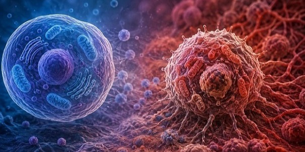
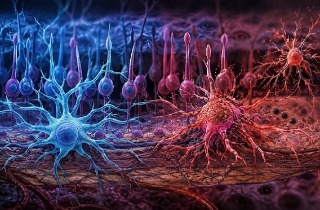
DN2 cells were enriched among people with high autoantibody levels and displayed increased TLR7 signaling, oxidative stress and antibody isotype switching. These biological changes could effectively place the cells into a highly activated state favoring production of abnormal antibodies.
COVID-19 Immune Cells Look Remarkably Like Lupus Cells
Perhaps the most concerning discovery was how closely COVID-associated DN2 cells resembled DN2 cells previously implicated in systemic lupus erythematosus.
Lupus is a chronic autoimmune disease in which the immune system attacks healthy tissues, potentially damaging the skin, joints, kidneys, blood vessels and other organs.
Researchers identified two important regulatory proteins, T-bet and XBP1, helping control the abnormal DN2 response. Their integrated genetic analysis also found that DN2 cells showed the strongest enrichment for inherited autoimmune-disease risk among the B-cell populations examined.
In other words, SARS-CoV-2 does not merely appear to cause random immune disruption. In susceptible individuals, infection may activate a recognizable biological program already associated with established autoimmune disease.
Another intriguing finding was that higher autoantibody abundance was inversely associated with neutralizing IgG antibodies. This suggests that some patients mounting stronger autoimmune antibody responses may simultaneously have less favorable levels of antibodies capable of neutralizing SARS-CoV-2.
Why the Findings Could Matter for Long COVID
The discovery could help scientists understand why some people develop persistent immune abnormalities or autoimmune complications following COVID-19 while others recover without them.
It also identifies possible therapeutic targets. If researchers can eventually interrupt excessive TLR7/8 signaling or the regulatory pathways involving T-bet and XBP1 without damaging normal immune defenses, it may become possible to reduce harmful post-infection autoimmunity.
However, the detailed molecular comparison involved a small group of selected female participants, meaning larger and more diverse studies will be needed before the findings can be translated into treatments.
Conclusions
The findings provide compelling mechanistic evidence that SARS-CoV-2 infection can activate DN2 B cells through pathways resembling those operating in lupus, potentially explaining how COVID-19 triggers autoantibody production and contributes to longer-term autoimmune complications in susceptible individuals.
The study findings were published in the peer reviewed journal: Immunity.
https://www.cell.com/immunity/abstract/S1074-7613(26)00324-9
Read Also:
https://www.thailandmedical.news/articles/long-covid




 Share
Share
 Tweet
Tweet
 Share
Share