German And Danish Scientists Using New Innovative X-Ray Imaging Shockingly Discover That SARS-CoV-2 Causes Extensive Heart Vascular Damage!
Source: Medical News - SARS-CoV-2 Heart Vascular Damage Dec 23, 2021 4 years, 6 months, 3 weeks, 1 day, 11 hours, 42 minutes ago
Scientist from the University of Göttingen-Germany, Hannover Medical School-Germany, Technical University of Denmark-Denmark and the University Medical Center of the Johannes Gutenberg University Mainz-Germany have in a new study using a new innovative x-ray imaging platform shockingly discovered that the SARS-CoV-2 coronavirus is able to cause extensive vascular damage to the heart! The study found significant changes in the heart muscle tissue of people who died from COVID-19.
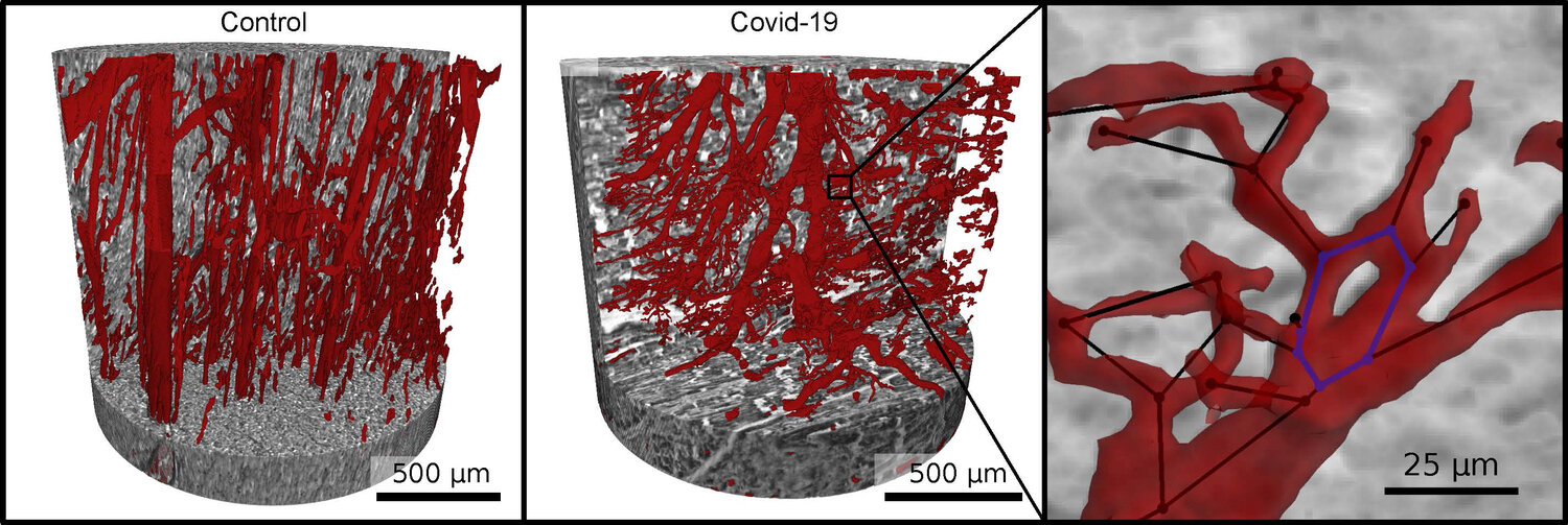 Vascular network (red) in healthy heart tissue (left) and in severe COVID-19 (right). Due to faulty reforming of the network as a result of COVID-19, numerous branches, splits and even loops develop in the capillaries, which can be analyzed mathematically. Credit: M. Reichardt, P. Møller Jensen, T. Salditt
Dear readers, we are appealing for your kind help and support this Christmas and the coming new year. We are desperately needing funds to sustain this website and all our research and community initiatives and need your help. Please donate. Thank You.
Vascular network (red) in healthy heart tissue (left) and in severe COVID-19 (right). Due to faulty reforming of the network as a result of COVID-19, numerous branches, splits and even loops develop in the capillaries, which can be analyzed mathematically. Credit: M. Reichardt, P. Møller Jensen, T. Salditt
Dear readers, we are appealing for your kind help and support this Christmas and the coming new year. We are desperately needing funds to sustain this website and all our research and community initiatives and need your help. Please donate. Thank You.
https://www.thailandmedical.news/p/sponsorship
So far damage to lung tissue has been the research focus in this area for some time and has now been thoroughly investigated but little is known about the degree of damage the SARS-CoV-2 coronavirus does to other organs, tissues and systems in the human host.
This is the first study that underpins the involvement of the heart in COVID-19 at the microscopic level for the first time by imaging and analyzing the affected tissue in the three dimensions.
The study team used a novel phase-contrast X-ray tomography to characterize the three-dimensional (3d) structure of cardiac tissue from patients who succumbed to COVID-19.
By extending conventional histopathological examination by a third dimension, the delicate pathological changes of the vascular system of severe Covid-19 progressions can be analyzed, fully quantified and compared to other types of viral myocarditis and controls. To this end, cardiac samples with a cross section of 3:5mm were scanned at a laboratory setup as well as at a parallel beam setup at a synchrotron radiation facility. The vascular network was segmented by a deep learning architecture suitable for 3d datasets (V-net), trained by sparse manual annotations.
Pathological alterations of vessels, concerning the variation of diameters and the amount of small holes, were observed, indicative of elevated occurrence of intussusceptive angiogenesis, also confirmed by high resolution cone beam X-ray tomography and scanning electron microscopy.
The study team implemented a fully automated analysis of the tissue structure in form of shape measures based on the structure tensor.
The corresponding distributions show that the histopathology of COVID-19 differs from both influenza and typical coxsackie virus myocarditis. The vascular damage to the heart was far more extensive to that ever seen before in any viral infections.
The study findings could also have implications as to why many &lsquo
;recovered’ COVID-19 patients tend to have heart issues weeks or months later although ignorant ‘experts’ are not linking it to COVID-19 or as to why the excess rates due to heart issues are ever increasing in the last two years.
The study findings were published in the peer reviewed journal: eLife.
https://elifesciences.org/articles/71359
The study team imaged the tissue architecture to a high-resolution using synchrotron radiation ie a particularly bright X-ray radiation and displayed it three-dimensionally.
In order to do this, the stud team used a special X-ray microscope that the University of Göttingen set up and operates at the German Electron Synchrotron DESY in Hamburg.
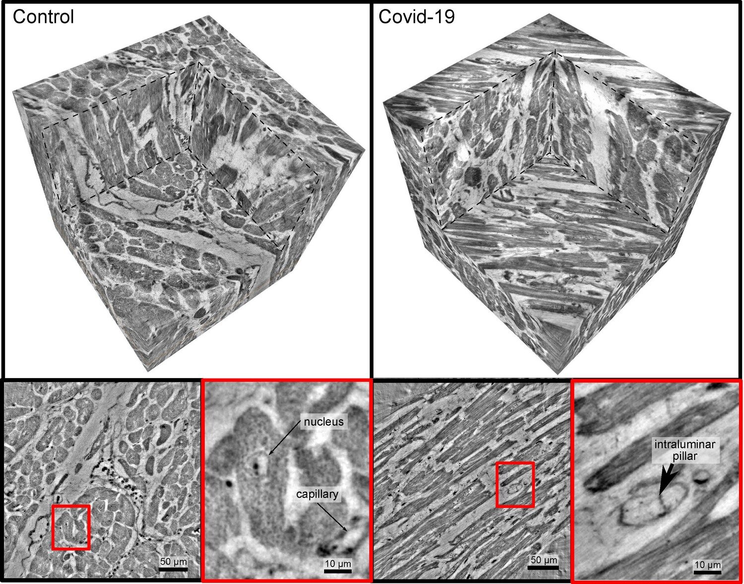 Healthy heart tissue (left) and in severe COVID-19 (right). While the blood vessels in the healthy case show an intact structure (bottom right), one can see cavities or pouches and tiny tubular structures forming in the fine blood vessels (capillaries) in the COVID-19 tissue sample, which indicate particular morphological changes. Credit: M Reichardt, T Salditt
The study team observed clear changes at the level of the capillaries (the tiny blood vessels) in the heart muscle tissue when they examined the effects there of the severe form of COVID-19 disease.
Interestingly in comparison with a healthy heart, X-ray imaging of tissues affected by severe disease, revealed a network full of splits, branches and loops which had been chaotically remodeled by the formation and splitting of new vessels.
These shocking changes are the first direct visual evidence of one of the main drivers of lung damage in COVID-19: a special kind of "intussusceptive angiogenes" (meaning new vessel formation) in the tissue.
Importantly in order to visualize the capillary network, the vessels in the three-dimensional volume first had to be identified using machine learning methods.
This required the study team to painstakingly, manually label the image data.
First author, Dr Marius Reichardt from the University of Göttingen told Thailand
Medical News, "To speed up image processing, we therefore also automatically broke the tissue architecture down into its local symmetrical features and then compared them. The parameters obtained from this then showed a completely different quality compared to healthy tissue, or even to diseases such as severe influenza or common myocarditis."
Study leaders Professor Dr Tim Salditt from the University of Göttingen and Professor Dr Danny Jonigk from t Hannover Medical School further explained, “There is a very special feature of this study ie in contrast to the vascular architecture, the required data quality could be achieved using a small X-ray source in the laboratory of the University of Göttingen. In principle, this means it could also be done in any clinic to support pathologists with routine diagnostics. Such health screening involving the heart is going to be very critical considering the damage that the novel coronavirus is causing.”
The study team plans to further expand in the future the approach of converting the characteristic tissue patterns into abstract mathematical values in order to develop automated tools for diagnostics, again by further developing laboratory X-ray imaging and validating it with data from synchrotron radiation.
Dear readers, we are appealing for your kind help and support this Christmas and the coming new year. We are desperately needing funds to sustain this website and all our research and community initiatives and need your help. Please donate. Thank You.
https://www.thailandmedical.news/p/sponsorship
For the latest
SARS-CoV-2 Research, keep on logging to Thailand Medical News.


