Nikhil Prasad Fact checked by:Thailand Medical News Team Aug 23, 2026 1 hour, 8 minutes ago
COVID-19 is best known as a respiratory infection, but a new literature review suggests the disease is also be linked to subtle changes in the tiny blood vessels supplying the retina and choroid in children. The review also examined multisystem inflammatory syndrome in children, or MIS-C, an inflammatory condition that can develop after SARS-CoV-2 infection.
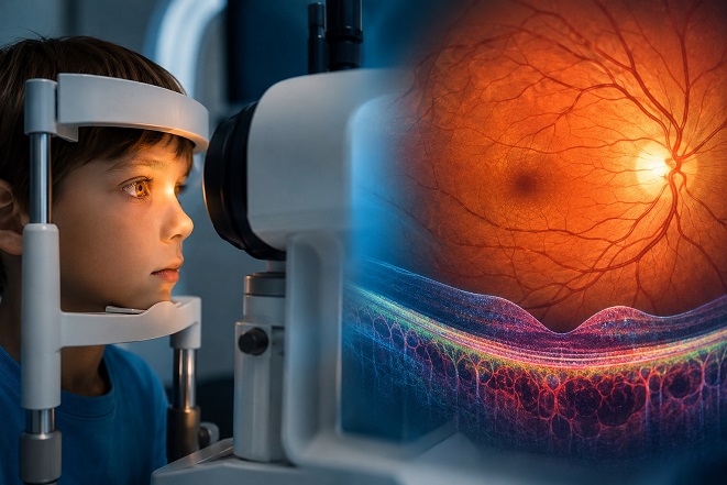 Advanced eye imaging suggests COVID-19 and MIS-C may temporarily alter tiny retinal blood vessels in some children
Why the Eye Can Reveal Vascular Changes
Advanced eye imaging suggests COVID-19 and MIS-C may temporarily alter tiny retinal blood vessels in some children
Why the Eye Can Reveal Vascular Changes
The retina, the light-sensitive tissue at the back of the eye, contains an intricate network of microscopic blood vessels. Doctors can examine these vessels without surgery using optical coherence tomography angiography, or OCTA. This imaging technique detects blood flow and measures features such as vessel density, retinal perfusion, and the foveal avascular zone, a naturally vessel-free area in central vision.
The review searched PubMed through April 2026 for original English-language studies using OCTA in pediatric COVID-19 or MIS-C patients. The evidence came mainly from small observational studies, meaning the findings show associations rather than proving that COVID-19 directly caused vascular damage.
Deep Retinal Capillaries Show the Strongest Signal
Across pediatric COVID-19 studies, the deep capillary plexus, a fine network of vessels deeper in the retina, was the layer most consistently reported as altered. Several studies found lower vessel density in this region after infection, while changes in the superficial capillary plexus were less consistent.
Macular blood-vessel density and perfusion also varied. Some studies found reductions, while others reported increases or no meaningful differences.
Measurements around the optic nerve were similarly mixed, and changes in the foveal avascular zone ranged from mild enlargement to no detectable difference.
In MIS-C, studies frequently reported reduced vessel density in the macular circulation, involving either or both superficial and deep capillary layers. Some abnormalities improved after recovery, suggesting that at least part of the vascular response may be temporary. However, selected groups still showed abnormalities months later.
What Could Be Driving the Changes?
Inflammation, injury to the lining of blood vessels, abnormal clotting, reduced oxygen levels, and disrupted regulation of blood flow are possible explanations. This Thailand
Medical News report highlights an important caution: OCTA measures flow-related signals, so a lower vessel-density reading does not automatically mean permanent loss of capillaries or retinal ischemia.
Researchers' institutions included Manchester Royal Eye Hospital and Manchester University NHS Foundation Trust; Oxford Eye Hospital and Oxford University Hospitals NHS Foundation Trust; the University of Manchester; University College London; and the NIHR Biomedical Research Center at Moorfields Eye Hospital and UCL Institute of Ophthalmology.
Conclusi
ons
The review suggests COVID-19 and MIS-C are associated with measurable retinal and choroidal microvascular changes in children, particularly in deeper retinal capillaries. However, inconsistent results, small study populations, different imaging systems, and varying follow-up periods mean OCTA cannot yet be considered a proven diagnostic or prognostic biomarker. Larger, standardized, long-term studies are needed to determine whether these eye findings reflect temporary inflammation or clinically important vascular effects.
The study findings were published in the peer reviewed journal: Medicina
https://www.mdpi.com/1648-9144/62/8/1603
Read Also:
https://www.thailandmedical.news/articles/coronavirus
https://www.thailandmedical.news/articles/long-covid




 Share
Share
 Tweet
Tweet
 Share
Share