UK Study Shows SARS-CoV-2 Causes Prolonged Change To Airway Immune Landscape With Evidence Of Cell Death Due To Ongoing Activation Of Cytotoxic T Cells!
Source: Post-COVID-19 Jan 27, 2022 4 years, 4 months, 3 weeks, 3 days, 19 hours, 48 minutes ago
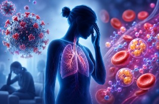
A new study led by researcher from National Heart and Lung Institute, Imperial College London-UK has alarmingly found that SARS-CoV-2 causes a prolonged change to the airway immune landscape in those with persistent lung disease, with evidence of cell death and tissue repair linked to ongoing activation of cytotoxic T cells.
The study also involved researchers and physicians from Westminster Hospital, Royal Brompton and Harefield Hospitals, Asthma UK Centre for Allergic Mechanisms of Asthma and the Margaret Turner-Warwick Centre for Fibrosing Lung Diseases.
Many patients hospitalized with acute COVID-19 often suffer respiratory symptoms that persist for many months.
The study team delineated the immune-proteomic landscape in the airway and peripheral blood of healthy controls and post-COVID-19 patients 3 to 6 months after hospital discharge.
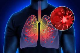
Post-COVID-19 patients showed abnormal airway (but not plasma) proteomes, with elevated concentration of proteins associated with apoptosis, tissue repair and epithelial injury versus healthy individuals.
Alarmingly, increased numbers of cytotoxic lymphocytes were observed in individuals with greater airway dysfunction, while increased B cell numbers and altered monocyte subsets were associated with more widespread lung abnormalities.
However, one-year follow-up of some post-COVID-19 patients indicated that these abnormalities resolved over time.
The study findings showed that SARS-CoV-2 infection causes a prolonged change to the airway immune landscape in those with persistent lung disease, with evidence of cell death and tissue repair linked to ongoing activation of cytotoxic T cells.
The study findings were published in the peer reviewed journal: Immunity.
https://www.cell.com/immunity/fulltext/S1074-7613(22)00046-2
The study team recruited 38 patients undergoing bronchoscopy for investigation of persistent respiratory abnormalities 3-6 months following acute SARS-CoV-2 infection. All patients had ongoing respiratory symptoms and/or radiological pulmonary abnormalities on computed tomography (CT). Peripheral blood and bronchoalveolar lavage (BAL) were obtained. The post-COVID19 cohort was stratified based on the level of respiratory support used during their initial hospitalization with acute COVID19, into moderate (no/minimal oxygen administered), severe (non-invasive ventilation) and very severe (invasive ventilation).
The key findings of the study were:
-Post-COVID19 airways, but not blood, show T cells, B cells macrophages and proteomic changes.
-Different post-COVID19 lung abnormalities relate to distinct immunological features.
-Increased BAL (bronchoalveolar lavage) cytotoxic T cells are linked to epithelial damage and airways disease.
-BAL myeloid and B cell numbers correlate with the degree of lung CT abnormality.
The acute immunological and inflammatory events that occur during human respiratory virus infections, including SARS-CoV-2, are relatively well described. In contrast, the immunolog
ical landscape of the human respiratory tract after recovery from acute viral infection is poorly understood. SARS-CoV-2 infection results in formation of long-lasting systemic immunological memory, with virus-specific antibodies and T cell responses still detectable in the majority of those infected at least 8 months post infection and higher titers seen in previously hospitalized individuals.
https://search.bvsalud.org/global-literature-on-novel-coronavirus-2019-ncov/resource/fr/covidwho-1309798
Circulating lymphocyte counts and the function and frequency of monocytes are also reduced during acute disease, but they appear to return to normal shortly after resolution of acute disease.
https://pubmed.ncbi.nlm.nih.gov/32943497/
Likewise, plasma concentrations of inflammatory mediators such as IL-6 and CXCL10, that are highly elevated in acute disease, reduce as individuals recover
https://pubmed.ncbi.nlm.nih.gov/32838342/
Collectively, this suggests that systemic inflammatory and immune responses associated with acute disease severity resolve in line with recovery from the acute symptoms. It therefore remains unclear if the severity of inflammation during acute disease is associated with the persistent respiratory pathology seen in some SARS CoV-2 infected individuals months after infection, or if there is ongoing inflammation in these individuals.
This study examines the relationship between the immune system and respiratory pathology post-COVID19. The immune cell and proteomic composition of the airways and peripheral blood were analyzed in a group of previously hospitalized COVID-19 patients with persistent radiological abnormalities in their lungs more than 3 months post discharge.
In comparison to healthy individuals, the post-COVID-19 airway showed substantial increases in activated tissue resident memory CD8+ and CD4+ tissue-resident memory (Trm) cells, and an altered monocyte pool. The airway proteome was also distinct from that observed in healthy individuals, with elevation in proteins associated with ongoing cell death, loss of barrier integrity and immune cell recruitment. None of these airway abnormalities were reflected in the proteome or immune cells of the matched peripheral blood.
The scale of these alterations was not linked to the initial severity of disease while in hospital and were heterogenous; some individuals displaying heightened T cell responses associated with significant increases in CXCR3 chemokines in the airways linked to prolonged epithelial damage and extracellular matrix (ECM) dysregulation, while other individuals exhibited a return to relative airway homeostasis. Subsequent long-term follow-up also suggested that these changes to the airway landscape progressively return to normal.
.jpg) Graphical Abstract
Graphical Abstract
The study findings showed upregulation of proteins associated with ongoing cell death, epithelial damage and tissue repair in post-COVID19 airways. This correlated with the presence of increased numbers of activated tissue resident CD8 T cells. Preliminary evidence suggests this altered airway landscape does improve over the long term, with reductions in airway immune cell numbers 1-year post discharge.
The acute response to SARS-CoV-2 infection is characterized by widespread upregulation of circulating proteins including IFN pathway proteins, chemokines, cytotoxic proteins, and markers of epithelial damage. More severe disease is associated with increased inflammatory proteins (e.g. IL-6, TNF, GM-CSF, IL-1RN and IL-18).
A similar pattern of upregulated proteins, especially chemokines like CXCL10 and cytokines such as IL-6, is seen in the airways during acute COVID19. 3-6 months after SARS-CoV-2 infection however, despite the presence of ongoing respiratory morbidity, the plasma proteins differentially expressed during acute disease appear to have returned to similar concentrations to those seen in healthy controls.
In contrast, the post-COVID19 airways continue to display an abnormal proteome, with both distinct and shared features to that seen in acute disease. Proteins linked to inflammation feature less prominently than in acute COVID-19, whereas upregulation of proteins involved in epithelial damage and repair (e.g. the EGFR ligand AREG and the epithelial marker KRT19) persist. MMP-3, which regulates the extracellular matrix (ECM), was also differentially upregulated in the post-COVID19 airway. MMP3 and AREG are both upregulated after influenza A virus (IAV) infection in vivo in mice, and in vitro in human fibroblasts and epithelial cells; and both are linked to epithelial repair and fibrosis in the lungs.
Elevated LDH and albumin in the airways provide further evidence of ongoing cell death and damage to respiratory barrier integrity post-COVID-19. This observation is reinforced by the upregulation of a module of correlated proteins in the post-COVID-19 BAL whose individual members reflect epithelial damage (EPCAM, KRT19), cell death (CASP3) and epithelial repair (TGFA), but also suggest a connection between these processes and immune cell recruitment and survival (CXCL9-11, IL-7). Increased cell death within the airways correlates with the frequency of T cells, primarily CD8 Trm cells, and with heightened respiratory dysfunction.
In mouse models of severe acute respiratory virus infection, CD8 T cells are known to act as a double-edged sword. Although the cytotoxic molecules and cytokines they release are essential for clearing virus, they can also cause tissue damage and immunopathology.
While pre-existing virus specific CD8 Trm cells in the airways is thought to be protective against a re-encounter with the same virus, little is known about their role in long-term respiratory virus-related pathology, especially in humans. This is primarily due to the lack of relevant samples collected during the recovery period.
The post-COVID-19 data support the concept that sustained activation of CD8 Trm cells in the airways, long after recovery from acute disease, contributes to ongoing damage to the respiratory epithelium, resulting in airway disease.
The mechanism underlying increased Trm cells in the airways is unclear, although several studies have reported virus specific CD8 T cells in lung tissue up to a year post-infection.
While virus specific CD4 and CD8 T cells rapidly expand, and form Trm cells, following SARS-CoV-2 infection, these cells rapidly contract after resolution of acute disease, with CD8 Trm cells declining more rapidly than CD4 Trm.
The lungs of mice previously experienced IAV infection more robustly maintain CD8 Trm cells compared to uninfected lungs however, showing that severe infection promotes a pro-Trm niche. This fits with the observation that CD8 Trm cell numbers vary dependent on the proteins and extent of damage in the airways, and change longitudinally in the same individuals, while CD4 Trm cells remain relatively static.
In the post-COVID-19 patients, peripheral blood monocyte had normalized and did not correlate with markers of pulmonary dysfunction, but BAL intermediate monocytes were increased in patients with greater CT abnormalities. In humans, following inflammatory insults, monocytes are recruited to the airways to differentiate into new AMs. Severe viral infection can cause rapid depletion of the airway macrophage pool, and different subsets of monocytes contribute differentially to the replenishment of lung macrophages. Monocyte to macrophage transition is also more pronounced in chronic lung disease, with the newly generated monocyte-derived macrophages acting in a pro-fibrotic fashion. Increases in intermediate monocytes may therefore be indicative of heightened monocyte differentiation into airway macrophages, the numbers of which are increased in the post-COVID-19 airway compared to healthy controls.
Amplification of this process may then contribute to ongoing repair within the lungs. The progressive resolution of radiological abnormalities in the majority of post COVID-19 patients has been described, and within our study even the 3 patients with persistent respiratory abnormalities show improved CT and reduced airway immune cell infiltration.
This fits with the hypothesis that SARS-CoV-2 infection can result in organizing pneumonia, with subsequent changes reflecting ongoing epithelial damage and healing parenchyma rather than established fibrosis. Moreover, the involvement of the immune response in different aspects of ongoing respiratory disease post-COVID-19 suggests this recovery could be accelerated using immunomodulatory treatments.
For more on
Post-COVID-19 Medical Conditions, keep on logging to Thailand Medical News.


 Share
Share
 Tweet
Tweet
 Share
Share