Parkinson’s Disease Breakthrough Reveals How a Natural Brain Toxin Called Aminochrome Triggers Dangerous Protein Build-Up
Nikhil Prasad Fact checked by:Thailand Medical News Team Jun 12, 2026 1 hour, 24 minutes ago
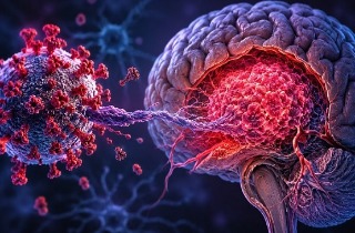
Medical News: Parkinson’s disease researchers have uncovered a potentially crucial mechanism that may help explain why brain cells gradually die in the disorder. The new study shows that aminochrome, a naturally occurring compound formed during dopamine metabolism, can disrupt one of the cell’s most important waste-disposal systems, causing harmful protein aggregates to accumulate inside neurons and ultimately contributing to cell death.
 Researchers discover that aminochrome, a natural byproduct of dopamine metabolism, may drive Parkinson’s disease by
blocking the brain cell’s waste-clearing machinery and promoting toxic protein accumulation
The research was conducted by scientists from the Centro de Investigación Austral Biotech and Universidad Santo Tomás in Santiago, Chile; the Molecular and Clinical Pharmacology unit at the Instituto de Ciencias Biomédicas, Faculty of Medicine, University of Chile; and Universidad Adolfo Ibáñez in Viña del Mar, Chile.
A Hidden Threat Produced Inside Brain Cells
Unlike many toxins linked to neurological diseases, aminochrome is not something that enters the body from the environment. It is produced naturally during the formation of neuromelanin, a dark pigment found in dopamine-producing neurons of the brain. Scientists have long suspected that aminochrome may play an important role in Parkinson’s disease because it can trigger oxidative stress, damage cellular structures, and interfere with normal neuronal functions.
Until now, however, researchers did not fully understand how aminochrome contributes to the formation of abnormal protein clumps, one of the hallmarks of Parkinson’s disease.
Protein aggregates, including those associated with Lewy bodies, are commonly found in the brains of people with Parkinson’s disease. These abnormal accumulations are believed to interfere with normal cellular activity and contribute to neurodegeneration.
The Cell’s Recycling System Comes Under Attack
Healthy cells continuously remove damaged proteins through sophisticated recycling systems. One of the most important of these systems is autophagy, a process in which cellular waste is enclosed inside structures called autophagosomes and then delivered to lysosomes, which act like microscopic recycling centers.
The new study found that aminochrome interferes with the critical fusion step between autophagosomes and lysosomes. When this fusion is disrupted, waste material cannot be properly broken down and removed.
Using laboratory-grown human neuronal cells that had been differentiated into dopamine-producing neurons, the researchers observed that aminochrome significantly reduced the ability of autophagosomes and lysosomes to merge. The effect was similar to that caused by vinblastine, a drug known to disrupt microtubules, the structural tracks that transport materials throughout cells.
The findings suggest that aminochrome damages the cellular transport network,
preventing waste-disposal components from reaching their destinations.
Protein Clumps Begin to Accumulate
As the recycling machinery became impaired, the researchers observed dramatic changes inside the cells.
One of the most striking findings was the accumulation of vimentin aggregates around the nucleus. Vimentin is a structural protein that often surrounds clusters of damaged proteins known as aggresomes. Cells exposed to aminochrome showed a six-fold increase in these vimentin-rich aggregates.
The team also detected large accumulations of ubiquitin, a molecule that normally labels damaged proteins for destruction. These ubiquitin deposits appeared not only around the nucleus but also inside the nucleus itself, an unexpected and potentially important discovery.
Even more concerning, aminochrome increased the overlap between ubiquitin and vimentin, indicating that damaged proteins were becoming trapped in growing aggregates that cells could no longer efficiently eliminate.
Evidence of Extensive Cellular Stress
The study revealed that aminochrome does not merely cause waste accumulation. It also appears to trigger a cellular emergency response.
Researchers found that aminochrome increased the expression of genes involved in autophagy and protein disposal. Levels of LC3β, a key autophagy-related gene, rose about five-fold, while ubiquitin gene expression increased approximately nine-fold. These changes suggest that neurons were desperately attempting to cope with the growing burden of damaged proteins.
Despite these efforts, many cells still died.
The investigators showed that aminochrome dramatically increased neuronal cell death. However, treatments that stimulate autophagy, including rapamycin and trehalose, partially reduced this toxicity. Trehalose appeared particularly effective, suggesting that strengthening cellular cleanup pathways could offer therapeutic benefits.
Why These Findings Matter
This
Medical News report highlights an emerging view of Parkinson’s disease in which the earliest events may involve damage to the cellular transport system rather than protein aggregation alone.
The researchers propose that aminochrome first destabilizes microtubules, disrupting intracellular transport. This then impairs the fusion of autophagosomes and lysosomes, causing damaged proteins to accumulate. Over time, these protein aggregates may overwhelm neurons and contribute to their degeneration.
Importantly, the discovery of ubiquitin aggregates inside the nucleus suggests that previously overlooked forms of protein toxicity may also be involved in Parkinson’s disease progression.
Conclusion
The study provides compelling evidence that aminochrome, a naturally occurring dopamine-derived toxin, can trigger a cascade of damaging events inside neurons. By disrupting microtubules and blocking the fusion of autophagosomes with lysosomes, aminochrome effectively cripples the cell’s ability to clear unwanted proteins. The resulting accumulation of vimentin and ubiquitin aggregates, including unusual deposits within the nucleus, points to a complex process of cellular dysfunction that may play a major role in Parkinson’s disease. These findings not only improve understanding of how neuronal death occurs but also identify promising therapeutic targets. Strategies aimed at stabilizing microtubules, restoring autophagy, improving protein clearance, and preventing toxic protein accumulation could potentially slow disease progression and protect vulnerable dopamine-producing neurons. Further studies will be needed to determine whether these mechanisms operate similarly in patients and how they can be translated into future treatments.
The study findings were published in the peer reviewed journal: Antioxidants.
https://www.mdpi.com/2076-3921/15/6/739
For the latest on Parkinson’s Disease, keep on logging to Thailand
Medical News.
Read Also:
https://www.thailandmedical.news/articles/alzheimer,-dementia-


 Share
Share
 Tweet
Tweet
 Share
Share