Efferocytosis of Virus-Containing Apoptotic Bodies Allows SARS-CoV-2 to Enter Low-ACE2 Macrophages and Trigger Inflammation
Nikhil Prasad Fact checked by:Thailand Medical News Team Jun 17, 2026 1 hour, 18 minutes ago
Medical News: A groundbreaking new study has uncovered an unexpected mechanism that may help explain how SARS-CoV-2 gains access to immune cells that were previously thought to be relatively resistant to infection. The research reveals that the virus can exploit a natural cellular cleanup process known as efferocytosis to enter macrophages and trigger the intense inflammatory responses associated with severe COVID-19.
 Researchers discover that SARS-CoV-2 can hide inside apoptotic bodies to enter macrophages and drive
severe inflammatory responses
The study was conducted by researchers from the Department of Biochemistry and Chemistry and the Research Centre for Extracellular Vesicles at La Trobe University, the La Trobe Institute for Molecular Science, The Walter and Eliza Hall Institute of Medical Research (WEHI), the Department of Medical Biology at the University of Melbourne, the Bio21 Molecular Science and Biotechnology Institute at the University of Melbourne, the VIB-UGent Center for Inflammation Research and Ghent University in Belgium, and Aston University in the United Kingdom.
Solving a Long-Standing COVID-19 Mystery
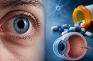
Since the beginning of the pandemic, scientists have known that SARS-CoV-2 primarily enters cells through the ACE2 receptor. However, a puzzling observation repeatedly emerged from studies of patients with severe COVID-19.
Macrophages, which are key immune cells involved in inflammation, were often found carrying viral material despite having very low levels of ACE2. This raised a critical question: how was the virus entering these cells?
The new study provides a compelling answer. Researchers discovered that when SARS-CoV-2 infects lung epithelial cells, many of these cells eventually undergo apoptosis, a programmed form of cell death. During this process, the dying cells fragment into small membrane-bound structures called apoptotic bodies.
Importantly, the researchers found that these apoptotic bodies were not empty cellular debris. Instead, many contained intact and infectious SARS-CoV-2 particles.
Viral Hitchhiking Inside Cellular Debris
Detailed laboratory analyses showed that apoptotic bodies released from infected cells carried viral spike proteins, nucleocapsid proteins, viral RNA, and even infectious virus particles capable of spreading infection.
The researchers observed that these virus-containing apoptotic bodies were readily engulfed by macrophages through efferocytosis, a natural process by which immune cells remove dead and dying cells from the body.
Rather than protecting the body, this cleanup mechanism inadvertently provided SARS-CoV-2 with an alternative route into macrophages.
Notably, direct exposure of macrophages to free SARS-CoV-2 resulted in minimal infection. In contrast, macrophages that engulfed virus-containing apoptotic bodies showed clear evidence of viral entry and accumulation of viral genetic material.
Triggering Dangerous Inflammatory Responses
The most signifi
cant finding was what happened after macrophages consumed these infected apoptotic bodies.
The macrophages began producing large quantities of inflammatory molecules strongly linked to severe COVID-19, including TNF, IL-6, IL-1β, IL-8, and IL-10. These cytokines are among the key drivers of the excessive immune activation commonly referred to as a cytokine storm.
Further investigations revealed activation of major inflammatory pathways, including inflammasome signaling and NF-κB signaling. These pathways are known to amplify immune responses and contribute to tissue injury in severe viral infections.
This
Medical News report highlights that the findings may help explain why macrophage-driven inflammation becomes such a dominant feature of severe COVID-19 despite the relatively low expression of ACE2 in many macrophage populations.
The researchers also found evidence of this phenomenon in infected mice. Significant numbers of apoptotic bodies were detected in the lungs, and many contained viral proteins. Their presence was associated with increased macrophage infection, elevated inflammatory cytokine production, and worsening lung pathology.
Potential New Therapeutic Opportunities
The team then searched for ways to interrupt this newly identified pathway. Through extensive screening, they discovered that several T-type calcium channel blockers could suppress the formation of apoptotic bodies. One drug in particular, mibefradil, significantly reduced the production of virus-containing apoptotic bodies without directly affecting viral replication.
When apoptotic body formation was reduced, macrophages showed less viral uptake, improved survival, and substantially lower inflammatory cytokine production.
In mouse models of severe COVID-19, treatment with mibefradil reduced the number of apoptotic bodies in the lungs, lowered viral burden, decreased inflammatory cell infiltration, and improved lung tissue damage.
The findings suggest that targeting apoptotic body formation may represent a completely new therapeutic strategy aimed not only at limiting viral spread but also at reducing the excessive inflammation that contributes to severe disease.
Conclusions
This study identifies a previously unrecognized mechanism by which SARS-CoV-2 can gain access to macrophages that express little or no ACE2. By hiding within apoptotic bodies released from dying infected cells, the virus effectively exploits the body's normal cellular clearance system to enter immune cells and provoke powerful inflammatory responses. The findings offer important new insights into COVID-19 pathogenesis and help explain how macrophage-driven cytokine storms develop during severe disease. They also highlight apoptotic body formation and efferocytosis-associated viral transmission as promising new targets for future therapeutic interventions against COVID-19 and potentially other viral infections that induce extensive cell death.
The study findings were published in the peer reviewed journal: Nature Communications.
https://www.nature.com/articles/s41467-026-74363-8
For the latest COVID-19 news, keep on logging to Thailand
Medical News.
Read Also:
https://www.thailandmedical.news/articles/coronavirus
https://www.thailandmedical.news/articles/long-covid


 Share
Share
 Tweet
Tweet
 Share
Share