Non-Human Primate Study Reveals How COVID-19 Disrupts Brain Energy and Immune Balance
Nikhil Prasad Fact checked by:Thailand Medical News Team Aug 10, 2025 11 months, 1 week, 21 hours ago
Medical News: Persistent Brain Changes After Infection
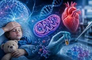
A new study by researchers from the Tulane National Primate Research Center, Tulane University School of Medicine, and biotechnology company Gigantest has shed light on how COVID-19 can cause long-lasting effects on the brain. Using African green monkeys, which closely mimic human responses to SARS-CoV-2, scientists tracked the impact of the virus 18 weeks after infection. While some signs of early brain injury—such as dying neurons and tiny brain bleeds—had improved, the researchers found that brain inflammation, changes in brain metabolism, and immune system disruptions were still present. This
Medical News report highlights how these changes could help explain the lingering neurological symptoms many people experience after COVID-19, often referred to as long COVID or neuroPASC.
 Non-Human Primate Study Reveals How COVID-19 Disrupts Brain Energy and Immune Balance
Inflammation That Refuses to Fade
Non-Human Primate Study Reveals How COVID-19 Disrupts Brain Energy and Immune Balance
Inflammation That Refuses to Fade
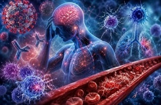
Even after the virus was no longer detectable in the brain, immune cells called microglia and support cells known as astrocytes remained in a state of overactivation. These cells clustered around blood vessels and lost their normal structure, suggesting ongoing damage to the blood-brain barrier—the protective shield between the brain and the bloodstream. The study also found increased numbers and size of microglial nodules, especially in the brainstem and cerebellum, indicating areas of concentrated inflammation.
Disrupted Oxygen and Energy Use
Interestingly, although the animals showed normal blood oxygen levels most of the time, their brain blood vessels continued to produce high levels of hypoxia-inducible factor-1α (HIF-1α). This protein is normally triggered by low oxygen but can also be activated by inflammation and high blood sugar. Indeed, the infected monkeys developed persistent increases in blood glucose, hinting at broader metabolic disruptions.
Metabolic Shifts in Brain Fluid
Cerebrospinal fluid analysis revealed significant changes in key molecules that power brain cells. Several important intermediates of the tricarboxylic acid (TCA) cycle—a core energy pathway—were out of balance. There was also a depletion of branched-chain amino acids, as well as anti-inflammatory molecules like adenosine and cortisol. These shifts are consistent with an energy metabolism rewired to support chronic inflammation rather than normal brain activity.
Changes in Taste and Thinking
Behavior tests showed that infected monkeys were more willing to eat bitter-tasting food, suggesting altered taste perception, and performed worse in a puzzle-solving task. They spent less time engaged and showed fewer attempts at problem-solving. These behavioral changes matched the degree of brain inflammation and microvascular damage, reinforcing the link between physical brain changes and functional decline.
Why This Matters for Long CO
VID
The study demonstrates that COVID-19 can leave a lasting footprint in the brain—altering immune activity, disrupting energy metabolism, and impairing cognitive function—months after the initial illness. The researchers warn that such changes could accelerate brain aging and increase the risk of neurodegenerative diseases. They stress the importance of monitoring and addressing brain health in COVID-19 survivors, even in younger individuals and those who did not have severe symptoms during the acute phase.
The study findings were published on a preprint server and are currently being peer reviewed.
https://www.researchsquare.com/article/rs-7134258/v1
For the latest COVID-19 news, keep on logging to Thailand
Medical News.
Read Also:
https://www.thailandmedical.news/news/sars-cov-2-proteins-found-to-damage-brain-cells-and-trigger-inflammation
https://www.thailandmedical.news/news/new-concerns-about-bone-health-after-covid-19
https://www.thailandmedical.news/news/covid-19-causes-skeletal-muscle-and-mitochondrial-damage-that-contributes-to-myalgic-encephalomyelitis-chronic-fatigue-syndrome


 Share
Share
 Tweet
Tweet
 Share
Share