Doctors Warn That COVID-19 Could Possibly Cause Unruptured Sinus of Valsalva Aneurysm
Nikhil Prasad Fact checked by:Thailand Medical News Team May 09, 2025 1 year, 3 weeks, 2 days, 15 hours, 24 minutes ago
Medical News: Medical experts are raising concerns over a potentially serious cardiovascular complication linked to COVID-19—unruptured sinus of Valsalva aneurysm (SVA). This rare but dangerous condition has now emerged in a new case report involving a post-COVID patient, prompting cardiologists to reevaluate the virus’s lingering effects on the heart and vascular system.
 Doctors Warn That COVID-19 Could Possibly Cause Unruptured Sinus of Valsalva Aneurysm
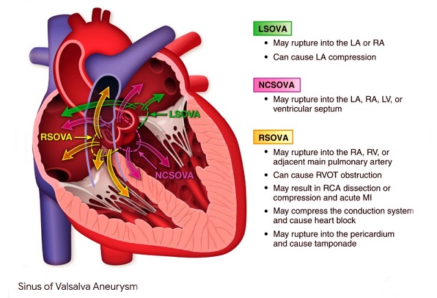
An unruptured sinus of Valsalva aneurysm (SVA) is a bulge in the aortic root, specifically the sinus of Valsalva, that hasn’t yet ruptured into a nearby heart chamber or blood vessel. SVAs are often congenital or acquired, and while unruptured SVAs can be asymptomatic, they can also cause complications like coronary artery compression, leading to symptoms like chest pain and shortness of breath. Unruptured SVAs are rare cardiac lesions that arise due to congenital or acquired causes and can exert pressure—called a “mass effect”—on the heart’s structures, including coronary arteries, valves, and surrounding tissues.
Now, researchers from the Department of Cardiovascular Ultrasound and the Department of Cardiovascular Surgery at Zhongnan Hospital of Wuhan University, China, have reported a rare case that may suggest a link between SARS-CoV-2 infection and the development or exacerbation of this vascular anomaly. This
Medical News report looks at the findings and their implications.
A Case That Raises Red Flags
The subject of the case report was a 55-year-old man from central China who presented with chest tightness—a symptom that initially appeared shortly after recovering from a COVID-19 infection. Despite having a 10-year history of hypertension and a heavy smoking habit, the patient had otherwise been considered in stable health.
During his initial evaluation one year ago, transthoracic echocardiography (TTE) revealed a non-coronary SVA measuring 5.0 cm, but no signs of aortic regurgitation were present. As the aneurysm was stable and didn’t meet criteria for surgery at the time, the medical team recommended smoking cessation and regular follow-ups.
At his recent visit, the patient again reported episodes of chest discomfort, though his blood pressure remained well-controlled. He had, however, continued smoking. A repeat TTE yielded similar findings. But further tests, including coronary computed tomography angiography (CCTA), revealed a troubling development—a large thrombus had formed within the aneurysm, and signs of arterial calcification were evident.
Bloodwork revealed elevated triglycerides, uric acid, and D-dimer levels—markers that signal both atherosclerosis and abnormal clotting activity. A brain CT scan also indicated a small left-sided stroke, likely a result of embolism caused by the clot.
Surgical Intervention and Recovery
Due to the serious risk of the thrombus dislodging and causing systemic emboli
sm—potentially leading to stroke or organ damage—the patient was quickly admitted for surgery. Intraoperative transesophageal echocardiography (TEE) confirmed the presence of a large mural thrombus in the non-coronary SVA.
Surgeons removed the thrombus and reconstructed the affected sinus using a prosthetic patch and autologous tissue. Postoperative imaging confirmed that the heart valves remained functional and no residual aneurysm was present. Remarkably, the patient made a full recovery and had quit smoking by the six-month follow-up. His repeat TTE showed normal heart structure and function.
Imaging Limitations and Diagnostic Challenges
This case also highlighted significant diagnostic challenges. TTE, although widely used as a first-line imaging tool for heart issues, failed to detect the thrombus due to calcification obscuring the view. It was only through advanced imaging—CCTA and intraoperative TEE—that clinicians could fully assess the situation.
The authors recommend using a combination of imaging modalities when managing SVAs to avoid missing critical findings. TEE and CCTA offer higher resolution and better visualization, especially when vessel calcification or acoustic limitations obscure results on standard ultrasound.
Could COVID-19 Be the Culprit?
While the exact cause of the aneurysm and subsequent thrombus formation cannot be definitively pinned on COVID-19, the researchers suggest the virus might have played a role.
The patient's prior infection with SARS-CoV-2, combined with his other risk factors—hypertension, smoking, and elevated blood lipids—could have triggered endothelial inflammation or “endotheliitis.” This inflammation, possibly accompanied by a COVID-induced cytokine storm, may have led to coagulopathy and impaired fibrinolysis, promoting clot formation in the weakened arterial wall.
Only a few cases in medical literature have proposed links between COVID-19 and vascular anomalies like SVA, but this case strengthens the need for further investigation.
Understanding Sinus of Valsalva Aneurysm
The sinus of Valsalva is one of three pouch-like structures at the base of the aorta. An aneurysm in this region often arises due to congenital defects but can also result from trauma, atherosclerosis, or infections—such as bacterial endocarditis, tuberculosis, and possibly COVID-19.
Unruptured SVAs often remain silent and are found incidentally during imaging for other reasons. When symptoms do occur, they can include chest pain, breathlessness, or palpitations—especially if the aneurysm begins to compress adjacent heart structures. The most feared complication is rupture, which can result in life-threatening internal bleeding.
Mural thrombi in SVAs pose their own risks. They can break free and travel to the brain or other organs, causing strokes or embolic events.
Why This Case Matters
This case is important for several reasons. It underscores the necessity of advanced imaging in detecting vascular complications that may not be visible on standard echocardiography. It also brings to light the possibility that COVID-19 might exacerbate or even cause rare vascular conditions in predisposed individuals.
Furthermore, it reminds clinicians to be vigilant in post-COVID patients who present with unexplained cardiac symptoms—even those with no history of major heart disease. Inflammatory and coagulative abnormalities induced by the virus may set the stage for complex vascular pathology.
Conclusion
The findings from this unique case provide compelling insights into how a COVID-19 infection might indirectly contribute to the formation or progression of rare vascular conditions like unruptured sinus of Valsalva aneurysms. While further research is necessary to confirm a causal link, the study adds to growing concerns about the long-term cardiovascular consequences of the virus.
Doctors should be aware of the limitations of standard imaging techniques like transthoracic echocardiography in evaluating such aneurysms and consider employing complementary tools such as coronary computed tomography angiography and transesophageal echocardiography, especially when symptoms persist or complications are suspected. Intraoperative TEE, in particular, was invaluable in guiding the surgical repair in this case and ensuring successful outcomes. Clinicians must also consider underlying coagulopathic states in post-COVID patients, especially when thrombus formation is evident.
Ultimately, while the patient in this case recovered fully and avoided long-term complications, his experience highlights the need for increased surveillance, multidisciplinary imaging approaches, and ongoing study into the cardiovascular aftereffects of COVID-19. For the general public, the takeaway is clear—COVID-19 can impact the body in unexpected ways long after the initial infection has passed, and new or recurring symptoms should never be ignored.
The study findings were published in the peer-reviewed journal: European Heart Journal – Case Reports.
https://academic.oup.com/ehjcr/article/9/5/ytaf201/8119850
For the latest COVID-19 News, keep on logging to Thailand
Medical News.
Read Also:
https://www.thailandmedical.news/news/case-study-highlights-that-covid-19-can-cause-massive-aortic-rupture
https://www.thailandmedical.news/news/covid-19-news-stanford-university-study-alarmingly-shows-that-sars-cov-2-infection-promotes-abdominal-aortic-aneurysm-progression
https://www.thailandmedical.news/news/utah-study-reveals-that-covid-19-and-influenza-increase-risk-of-acute-aortic-syndrome
https://www.thailandmedical.news/articles/coronavirus
https://www.thailandmedical.news/pages/thailand_doctors_listings


 Share
Share
 Tweet
Tweet
 Share
Share