Nikhil Prasad Fact checked by:Thailand Medical News Team Sep 13, 2026 1 hour, 14 minutes ago
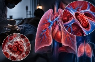
A newly identified population of dysfunctional immune cells may persist for months after acute COVID-19, offering fresh evidence that SARS-CoV-2 can leave measurable changes in the immune system well beyond the initial infection.
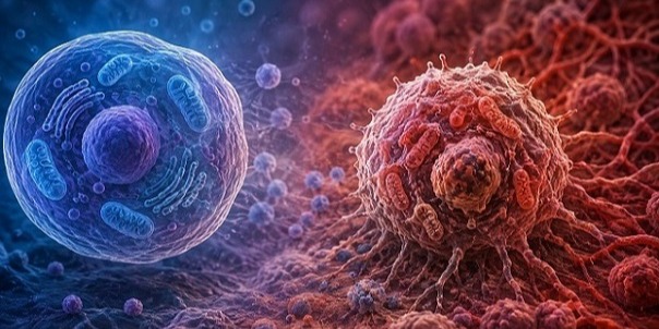
Researchers found that COVID-19 was associated with an expansion of unusual monocytes—white blood cells that help coordinate inflammation, destroy microbes, and develop into other immune cells. The cells showed a strikingly weak inflammatory response and remained abnormal for at least three months after diagnosis.
 COVID-19 was linked to dysfunctional monocytes that remained abnormal for at least three months after infection.
Researchers track immune changes over time
The research involved scientists from the Burnet Institute; RMIT University; The Alfred Hospital and Monash University; Monash University’s School of Population Health and Preventive Medicine; the University of Melbourne’s School of Population and Global Health; the Peter Doherty Institute for Infection and Immunity at the University of Melbourne; and Monash University’s Department of Microbiology.
The team analyzed stored blood samples from 42 people with PCR-confirmed COVID-19 and 40 uninfected controls. Participants with COVID-19 had a median age of 43 and were unvaccinated, with infections occurring in 2020 before Alpha, Delta, or Omicron circulated in Australia. About 59.5% were not hospitalized, 11.9% were hospitalized without requiring oxygen, and 28.6% were hospitalized and required oxygen.
Samples were examined during acute infection and, where available, about one month and three months after diagnosis.
An unusual monocyte population emerges
Using conventional markers, researchers detected a CD14lowCD16- monocyte population that was significantly expanded during acute COVID-19. The increase occurred in both mild and hospitalized cases but was greater in severe disease.
Because CD14 and CD16 can be shed from activated monocytes and potentially distort measurements, the investigators also used a five-receptor identification method. That independent approach confirmed the unusual population, which they named infection-associated monocytes, or IAM.
In this
Thailand Medical News report, one of the most important findings is that IAM were not simply more numerous: they behaved differently. Their abundance was positively associated with blood levels of TNF and CXCL10, inflammatory signaling molecules, but not with C-reactive protein, viral load, soluble CD14, soluble CD163, or CCL2.
Immune cells became unusually unresponsive
When researchers challenged the cells with lipopolysaccharide, a bacterial component commonly used to provoke an immune response in laboratory experiments, monocytes from people with acute COVID-19 produced less IL-1β, TNF, and IL-6.
The dysfunction was especially clear in non-classical monocytes and IAM. After stimulation, 62% of non-classical monocytes from COVID-19 cases produced none of the measured inflammatory cytokines, compared
with 29% in controls. For IAM, the figures were 63% versus 29%. Reduced responses also occurred after other bacterial and viral receptor stimuli, suggesting the defect was not limited to one experimental trigger.
Abnormalities remained three months later
The IAM population remained significantly elevated at one and three months. Compared with controls, IAM proportions were 16.9 percentage points higher at one month and 8.8 points higher at three months. IL-1β remained reduced across monocyte subsets, while non-classical monocytes and IAM continued to show impaired responses to stimulation.
Researchers also observed elevated activation of CD4+ and CD8+ T cells and natural killer cells through three months, suggesting broader immune disruption after infection.
Conclusions
The findings suggest COVID-19 can produce persistent changes in innate immune cells, including expansion of a previously underrecognized, poorly responsive monocyte population. However, the small observational study cannot establish whether these abnormalities cause long COVID or other clinical outcomes, and studies using fresh whole blood are needed to confirm their significance.
The study findings were published in the peer reviewed journal: Frontiers in Immunology.
https://www.frontiersin.org/journals/immunology/articles/10.3389/fimmu.2026.1814210/full
Read Also:
https://www.thailandmedical.news/articles/coronavirus
https://www.thailandmedical.news/articles/long-covid




 Share
Share
 Tweet
Tweet
 Share
Share