Nikhil Prasad Fact checked by:Thailand Medical News Aug 23, 2026 1 month, 2 weeks, 1 day, 5 hours, 59 minutes ago
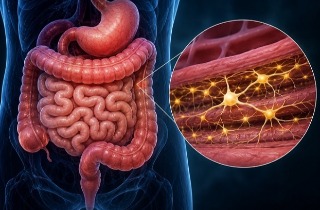
Diabetes may begin changing the nervous system inside the digestive tract before obvious stomach or bowel symptoms appear, according to a new animal study examining how prolonged high blood sugar affects nerve cells throughout the gut.
 Diabetes caused substantial NPY-related nerve changes across several digestive regions in an experimental pig model
The research focused on neuropeptide Y (NPY), a naturally occurring chemical messenger used by nerve cells. In the digestive system, NPY helps regulate movement of food, digestive secretions, water and electrolyte absorption, and blood flow. Because NPY generally slows intestinal activity and can narrow blood vessels, changes in its activity could potentially influence digestive problems associated with diabetes.
How the Study Was Conducted
The experiment involved 10 young female pigs, divided equally into diabetic and control groups. Diabetes was experimentally induced, producing sustained high blood sugar for six weeks. Tissue was then examined from the stomach, duodenum, jejunum, ileum, and descending colon.
Scientists specifically measured the proportion of gut nerve cells showing NPY. Pigs were selected because their digestive anatomy, intestinal organization, metabolism, and enteric nervous system—the network of nerves controlling the gut—share important similarities with humans.
The research institutions were the University of Warmia and Mazury in Olsztyn, Faculty of Veterinary Medicine, and the InLife Institute of Animal Reproduction and Food Research, Polish Academy of Sciences, Poland.
Major Changes Appeared Across the Gut
The strongest finding was that diabetes did not affect every part of the digestive nervous system equally. In this Thailand
Medical News report, the results show particularly notable changes in the myenteric plexus, the nerve network largely involved in controlling gut movement.
In the stomach, NPY-positive neurons increased from 1.93% in controls to 3.70% in diabetic animals. In the jejunum, they rose from 2.73% to 4.78%, while the ileum showed an increase from 3.18% to 5.09%.
The most dramatic changes appeared in the descending colon. NPY-positive neurons in its myenteric plexus increased from 2.89% to 6.12%, more than doubling. The inner submucosal plexus increased from 1.77% to 2.99%, while the outer submucosal plexus jumped from 0.84% to 2.23%, approximately a 2.65-fold increase.
Although increases were also recorded in the duodenum, they were not statistically significant after researchers adjusted for multiple comparisons. Overall, significant myenteric changes occurred in the stomach, jejunum, ileum, and descending colon, while significant submucosal changes were confined to the descending colon.
What the Findings Could Mean
Importantly, the pigs had no obvious diarrhea, constipation, appetite problems, or other clinically apparent digestive disturbances during the experiment. This suggests nerve-related biochemical changes may emerge before noticeable gast
rointestinal symptoms.
The researchers caution that increased NPY could represent a protective response helping nerve cells cope with metabolic stress, but altered NPY signaling might also contribute to slower gut movement, altered secretion, reduced local blood flow, and eventually diabetic gastrointestinal dysfunction.
Conclusion
These findings suggest sustained high blood sugar can reshape chemical signaling within gut nerves in highly specific locations, potentially providing an early biological mechanism linking diabetes with later digestive complications.
However, larger and longer studies are needed to establish whether these nerve changes protect the gut or ultimately contribute to disease.
The study findings were published in the peer reviewed International Journal of Molecular Sciences.
https://www.mdpi.com/1422-0067/27/17/7534
Read Also:
https://www.thailandmedical.news/articles/diabetes 



 Share
Share
 Tweet
Tweet
 Share
Share