Nikhil Prasad Fact checked by:Thailand Medical News Team Aug 16, 2026 1 month, 2 weeks, 2 hours, 38 minutes ago
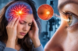
The blue light produced by digital screens may do more than cause temporary eye discomfort. New laboratory research suggests prolonged blue-light exposure can activate a destructive form of cell death in the cornea, potentially contributing to dry eye disease.
 Researchers found that sustained blue-light exposure can weaken corneal antioxidant defenses and trigger an iron-dependent
form of cell death associated with dry eye
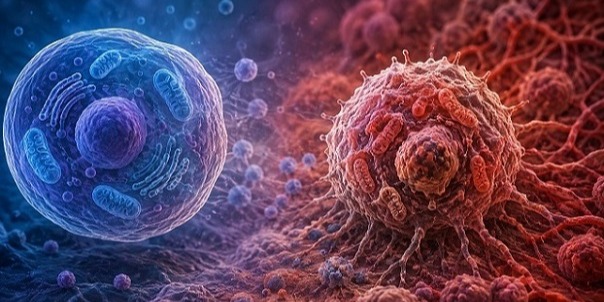
Researchers found that blue light activated the stress-related protein p53, which then weakened important antioxidant defenses inside corneal cells. The resulting damage involved ferroptosis, a form of cell death driven by iron accumulation and damaging oxidation.
What Is Blue Light Pollution?
Blue light is part of the high-energy visible light spectrum, generally covering wavelengths of about 400 to 500 nanometers. Modern lifestyles have greatly increased exposure to artificial sources of blue light, particularly smartphones, tablets, computer monitors, televisions and other digital displays. The researchers describe this increasingly widespread environmental exposure as blue light pollution.
Unlike occasional natural exposure, digital-device use can expose the eyes to artificial blue light repeatedly for many hours. Because the cornea forms the eye's outermost protective surface and directly encounters incoming light, researchers are increasingly interested in whether sustained exposure could affect corneal cells and contribute to ocular surface problems such as dry eye.
Researchers and Institutions
The research was conducted by Lingyu Zhang, Yiwen Qian, Jun Jin, Yu Zhang, Zhiliang Wang and Qingjian Li.
The researchers are from the Tianjin Key Laboratory of Retinal Functions and Diseases, Tianjin Branch of the National Clinical Research Center for Ocular Disease, Eye Institute and School of Optometry, Tianjin Medical University Eye Hospital, Tianjin, China; and the Department of Ophthalmology, Huashan Hospital, Fudan University, Shanghai, China.
Blue Light Gradually Damaged the Eye Surface
Researchers exposed mice to 460-nanometer blue light at 1,000 lux for eight hours daily for one, two or four weeks. Human corneal epithelial cells were separately exposed for 15 or 30 minutes.
Damage became progressively worse with longer exposure. The mice developed disruption of the corneal surface, poorer tear-film stability and increasing inflammatory-cell infiltration. After four weeks, the epithelial layer covering central and peripheral areas of the cornea became significantly thinner.
Importantly, overall corneal thickness and transparency remained largely preserved, suggesting that much of the observed injury was concentrated in the surface epithelial layer.
Cells Lost Important Antioxidant Defenses
Laboratory experiments showed that blue light reduced corneal-cell survival and caused cells to become trapped in the G1 stage of the cell cycle. Levels of CCND1 and CDK4, which help cells progress through this cycle, also fell. This could interfere with the cornea's ability to replace damaged surface cells.
;
In this Thailand
Medical News report, another major finding involved ferroptosis. Researchers detected increased ferrous iron and reactive oxygen species, unstable molecules capable of damaging cellular structures.
Levels of SLC7A11 and GPX4 also fell. These proteins are important components of the cellular antioxidant defense system. Meanwhile, COX2, a marker associated with ferroptosis, increased.
p53 Emerged as the Key Molecular Switch
RNA sequencing identified 901 genes significantly altered following blue-light exposure, including 579 that became more active and 322 that became less active. Analysis highlighted the p53 and ferroptosis pathways.
The researchers concluded that activated p53 suppresses SLC7A11, weakening the SLC7A11/GPX4 antioxidant system and making corneal cells more vulnerable to ferroptosis. When scientists experimentally reduced p53 activity, SLC7A11 and GPX4 recovered while COX2 decreased.
Experimental Eye Treatment Reduced Damage
Researchers then tested the p53 inhibitor pifithrin-alpha, or PFT-α, as a topical treatment in blue-light-exposed mice.
Treatment reduced corneal surface injury, improved tear-film stability and decreased inflammatory-cell infiltration. It also reduced inflammatory proteins IL-1β, IL-6 and TNF-α while restoring SLC7A11 and GPX4.
Conclusions
The findings suggest that sustained blue-light exposure can damage corneal epithelial cells through p53 activation, weakened antioxidant defenses, ferroptosis, inflammation and reduced cellular regeneration. Blocking p53 substantially protected the ocular surface in experimental models, highlighting a potentially important therapeutic pathway. However, these findings come from mice and cultured cells, and human clinical studies are still required before such treatments can be recommended for people experiencing screen-associated dry eye.
The study findings were published in the peer reviewed journal: Antioxidants.
https://www.mdpi.com/2076-3921/15/8/1014
Read Also:
https://www.thailandmedical.news/articles/ophthalmology-(eye-diseases)




 Share
Share
 Tweet
Tweet
 Share
Share