Nikhil Prasad Fact checked by:Thailand Medical News Team Jul 21, 2026 3 months, 2 days, 20 hours, 19 minutes ago
Medical News: For years, doctors have known that Myalgic Encephalomyelitis/Chronic Fatigue Syndrome (ME/CFS) and long COVID share many of the same disabling symptoms, including crushing fatigue, brain fog, memory problems, poor concentration, pain, sleep disturbances, and worsening symptoms after physical or mental activity. However, scientists have struggled to determine whether these illnesses affect the brain in the same way or through different biological mechanisms.
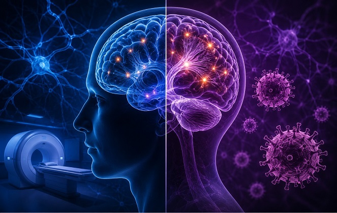 Advanced MRI scans reveal distinct microscopic brain changes that differentiate ME/CFS from long COVID while explaining
many shared neurological symptoms
Now, researchers from the National Center for Neuroimmunology and Emerging Diseases (NCNED), Griffith University, Gold Coast, Australia, have uncovered compelling evidence that while the two conditions share neurological changes, they also have distinct patterns of brain tissue damage.
Advanced MRI Technology Reveals Hidden Brain Changes
The research team recruited 37 people with ME/CFS, 19 individuals with long COVID, and 27 healthy volunteers. Every participant underwent sophisticated brain scans using a powerful 3-Tesla MRI system equipped with two advanced imaging techniques known as Diffusion Tensor Imaging (DTI) and Diffusion Kurtosis Imaging (DKI). These technologies can detect microscopic changes in brain tissue that conventional MRI scans cannot reveal.
The investigators compared the brain scans with clinical assessments measuring fatigue, quality of life, physical function, cognitive ability, and disability. As expected, both ME/CFS and long COVID patients reported substantially poorer health and greater disability than healthy volunteers, with people suffering from ME/CFS generally experiencing the greatest impairment.
Different Brain Regions Are Affected
The findings showed that people with ME/CFS had significant microscopic changes in several important brain regions, including the cingulum, supplementary motor area, and parts of the corpus callosum, the major nerve bridge connecting the brain's two hemispheres.
These areas help regulate thinking, memory, attention, emotional processing, movement planning, and communication between different parts of the brain. Damage or disruption in these regions could help explain why many patients struggle with persistent brain fog, slowed thinking, memory problems, and difficulty performing everyday activities.
Meanwhile, individuals with long COVID displayed a different pattern of abnormalities. Changes were found in the fusiform gyrus, precentral gyrus, superior corona radiata, and additional white matter pathways that are involved in visual processing, voluntary movement, attention, and communication between various brain regions.
Study Highlights a Major Discovery
Importantly, this
Medical News report describes the first study to directly compare ME/CFS and long COVID using both DTI and DKI brain imaging techniques in the same investigation.&
lt;br />
Although the standard DTI scans detected few differences between the two illnesses, the more advanced DKI method identified a significant difference in the left corona radiata, a major nerve fiber pathway that carries signals involved in movement, sensory processing, and higher brain functions.
Researchers believe this difference may reflect increased tissue complexity, altered cell density, or ongoing neuroinflammatory activity. The discovery suggests that DKI may provide a much more sensitive way of detecting subtle brain abnormalities that traditional MRI methods might miss.
Why These Findings Matter
The study also provides important clues about the biological basis of symptoms experienced by millions of patients worldwide.
In ME/CFS, the observed abnormalities could reflect altered myelin—the protective coating around nerve fibers—as well as changes in cellular density and nerve integrity. Reduced integrity within the corpus callosum may interfere with communication between the brain's hemispheres, potentially contributing to cognitive dysfunction, slower information processing, sensitivity to light and noise, and movement difficulties.
For long COVID, the abnormalities identified in regions involved in attention, motor control, and facial recognition may indicate persistent inflammation, changes in supporting brain cells, or altered white matter organization. These microscopic alterations may help explain ongoing fatigue, concentration problems, movement issues, and visual or cognitive symptoms reported by many patients months after their initial COVID-19 infection.
Although researchers observed several relationships between brain imaging findings and clinical symptoms, these associations were not statistically significant after rigorous correction for multiple testing, indicating that larger future studies will be needed to confirm these links.
Conclusion
This study provides some of the strongest evidence to date that both ME/CFS and long COVID involve genuine biological changes within the brain rather than symptoms without an identifiable neurological basis. At the same time, the research demonstrates that each illness follows its own distinctive pattern of microscopic brain alterations. The findings also suggest that advanced MRI techniques, particularly Diffusion Kurtosis Imaging, could become valuable tools for improving future research, identifying disease-specific changes, and eventually supporting more accurate diagnosis and targeted treatment strategies.
The study findings were published in the peer reviewed journal: Frontiers in Medicine.
https://www.frontiersin.org/journals/medicine/articles/10.3389/fmed.2026.1824498/full
For the latest on Long COVID, keep on logging to Thailand
Medical News.
Read Also:
https://www.thailandmedical.news/articles/long-covid




 Share
Share
 Tweet
Tweet
 Share
Share