BREAKING NEWS
For the latest on Thailand Medical Industry, Thailand Doctors, Thailand Medical Research, Thailand Hospitals, Thailand Wellness Initiatives and the latest Medical News
The human eye is a wonder of engineering. It consists of many different parts that work together to provide visual information to the brain, which then translates it into information that is useful to the body.
1. The cornea
The first step in this complex process occurs when light passes through the clear slightly convex cornea at the very front of the eye. This is the transparent part of the eyeball.
A thick white sheath called the sclera surrounds the rest of the eyeball. The cornea refracts light slightly. The narrow, liquid-filled space behind the cornea is called the aqueous humor. This drains through spaces at the medial corner of the eye, and is constantly renewed.
2. The iris
The iris is a colored diaphragm of thin circular and longitudinal muscle fibers just behind the cornea. It has an aperture in the center. This can expand or contract to let in more or less light, respectively, depending on the light in the surroundings.
This opening is called the pupil. Light passing through the cornea and the pupil falls on the anterior surface of the lens. The aqueous humor keeps the iris from sticking to the lens behind and the cornea in front.
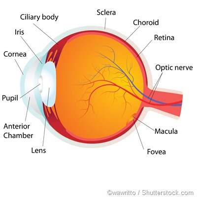
3. The lens
The lens is a clear crystalline globe which almost touches the posterior surface of the pupillary opening. The ciliary muscles are attached to the surface of the lens. The help the lens to change shape in order to focus.
As they contract, they cause the lens to become more round or long, so that the rays bend more or less, according to need. If the object focused on is far away, the lens needs to bend the light rays from it more sharply, to make them fall on the center of the retina, where vision is sharpest. For objects close-up, the lens becomes elongated so that light rays are bent less.
4. The posterior chamber
The refracted rays now pass through the jelly-like tissue that fills out the eyeball behind the lens. This part is called the posterior chamber. At the back, the eyeball is bounded by the choroid, a network of capillaries which nourishes all the structures of the eye.
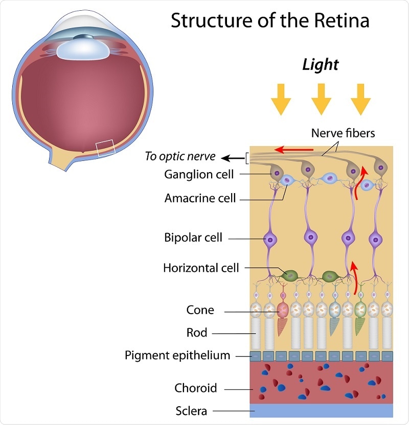
In front of it lies the retinal pigment epithelium, a layer of melanin-rich cells which supplies special nutrition to the sensory layer of the eye. The retina is nourished and renewed by the pigment epithelial cells.
5. The retina
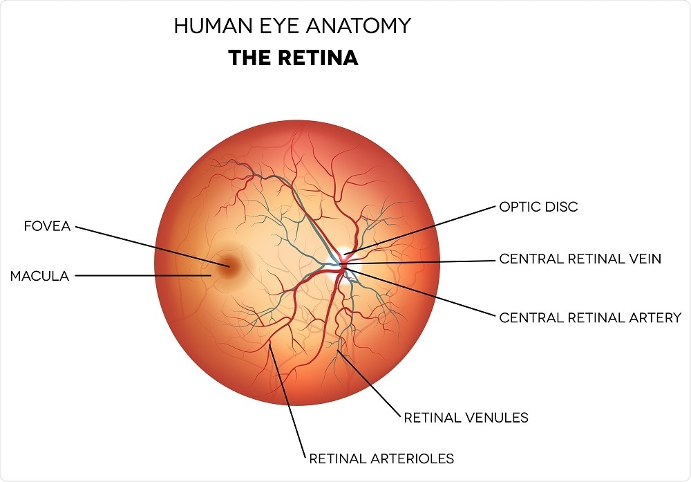
The retina is a multilayer membrane comprising a sensory photoreceptor array, a few layers of connecting neurons and an inner ganglion cell layer. The axons from the ganglion cells travel backward to pierce the retina and leave the eye through the optic nerve. There is a blind spot in the retina where the ganglion cells pass through.
The photoreceptors in the eye consist of rod and cone cells. The rods are found mostly in the peripheral part of the retina and are responsible for perception of light and dark, including shades of gray. They are more numerous than cones, and are very sensitive to light.
The cone cells are responsible for visual acuity and color vision, and millions of them are closely assembled in the central part of the retina, also called the macula. At the fovea, which is the central point of the macula, only cones are present, and normal vision uses this point to achieve sharp vision at maximum resolution.
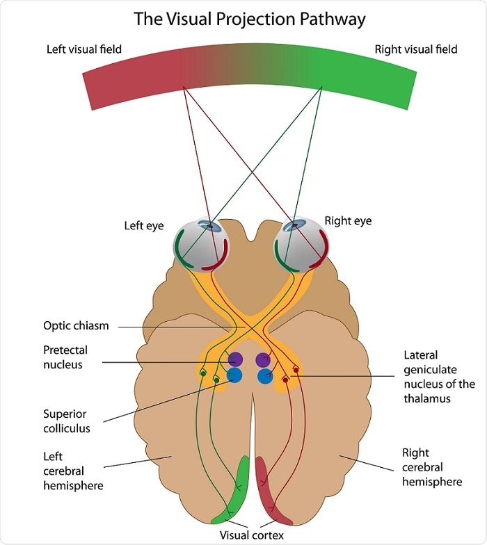
As the light rays fall on the photoreceptor cells, changes occur in the pigments they contain. This leads to bleaching of the pigments, and electrical impulses are generated. These are transmitted through a chain of neurons to the ganglion cells which carry the impulses to the visual cortex of the brain. There they are processed and the object is seen.
Each eye receives information from half of the visual field. Thus the middle parts of both fields overlap, and this leads to binocular vision. However, the difference in the peripheral parts of the left and right fields of vision lead to depth perception or three-dimensional vision. It helps in gauging distances accurately and estimating the depths and dimensions of objects.