

BREAKING NEWS
For the latest on Thailand Medical Industry, Thailand Doctors, Thailand Medical Research, Thailand Hospitals, Thailand Wellness Initiatives and the latest Medical News
Optical coherence tomography (OCT) is an imaging technique that uses light to capture 2D and 3D images up to a resolution of a micrometer (μm). It has many uses in medical imaging and research.
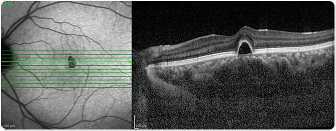
OCT is a high-resolution optical imaging technique that is based on the interference between the signal from the object being investigated (the sample) and a local reference signal. OCT can produce a cross-sectional image of an object in real time. For OCT, the axial resolution of the image is dependent on the optical source, which is an advantage when compared to other methods such as confocal microscopy. OCT can be used to gain very high-resolution images of the retina even with optical aberrations and when narrow light beams are used.
There are two main methods of OCT, time domain (TD)-OCT and spectral domain (SD)-OCT. Both have pros and cons and are varied in their procedure. SD-OCT is performed in two ways: spectrometer-based (SB), or using a tunable laser or a swept source (SS).
Time-domain optical coherence tomography
A TD-OCT setup uses an optical light source and an interferometer. A reference mirror and an optical splitter are used to produce a reference beam. A microscopy interface optics is used to convey light from the splitter to and from the object of interest and up to a processing unit. The processing unit performs the interference of light between the reference beam and the beam returned from the object of interest, and it also processes and analyzes the interference signal. The optical path difference (OPD) is obtained by subtracting the length of the reference path from the length of the object path. The OPD can be used to work out which layers within an object can be imaged.
OCT was invented by the addition of a transversal scanner to a low coherence interferometer to scan the beam over the object laterally. A cross-section image is obtained by taking adjacent scans of the object; this is also known as axial TD-OCT. Another method of performing TD-OCT is en face OCT, which involves taking one-dimensional reflectivity scans (T-scans), with the end image being comprised of many T-scans.
Spectral domain optical coherence tomography
SD-OCT refers to the spectral interrogation of the spectrum at the interferometer output. This can be done in two ways. The first involves using a combination of broadband optical source and the processing unit which utilizes a spectrometer. The second involves using a swept source (SS) and the processing unit which employs a photodetector. This is like the TD-OCT procedure, but instead of scanning the OPD the charges on the array are read in the spectrometer in SB-OCT or by tuning the frequency of the laser in SS-OCT.
SB-OCT is based on the demodulation output of the optical spectrum of the low coherence interferometer. The spectrum has peaks and troughs, the period of the modulation being proportional to the OPD in the interferometer. This relationship means the larger the OPD, the larger number of peaks in the spectrum. The camera used to image needs pixels small enough to be able to sample the peaks and troughs in the spectrum. In SB-OCT, the cameras used are at 20-70 kHz.
With SS-OCT the laser line needs to be narrower than the spectral distance between adjacent peaks for the photodetector to detect it when tuning the SS. This allows the photo-detected signal to take the shape of the channeled spectrum. This method translates the periodicity of the spectrum into peaks of differing frequency in relation to the OPD, which gives an A-scan. The A-scan is then used to produce an image.
There are several modes of OCT that can be used to image an object. They each have advantages and disadvantages that dictate which one is used in any given situation. These methods of OCT have been developed over many years and more research is going on to improve its efficiency. This will allow better images to be produced, which would be of great use to both researchers and the healthcare industry.