

BREAKING NEWS
For the latest on Thailand Medical Industry, Thailand Doctors, Thailand Medical Research, Thailand Hospitals, Thailand Wellness Initiatives and the latest Medical News
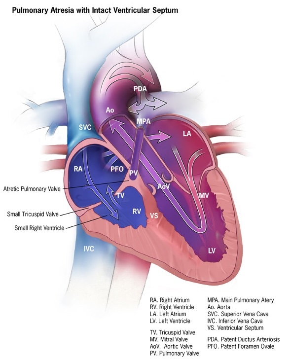
The term ‘pulmonary’ refers to the lungs and ‘atresia’ means missing, so pulmonary atresia is a congenital heart defect in which the pulmonary valve of the infant is missing. The pulmonary valve regulates the flow of blood from the right ventricle in the heart to the lungs, taking blood low in oxygen and passing it on to the lungs to make it oxygen rich.
In an infant born with pulmonary atresia, the pulmonary valve does not develop. Instead, a solid sheet of tissue exists where the valve should be. This effectively blocks the flow of blood. As a result of this birth defect, the infant suffers from a lack of oxygen-rich blood, which can be fatal. The condition often requires surgery soon after birth and is called a critical congenital heart defect.
Noticeable symptoms in a newborn with pulmonary atresia can become evident within a few hours of birth or they may be delayed for several days, depending on the type and severity of the condition. The symptoms could include:
A number of complications may arise from this condition. The baby will be at risk for seizures, strokes, or heart failure. The baby may also experience delayed growth and development, or infectious endocarditis. It is imperative that pulmonary atresia is identified as soon as possible for the baby to survive. If not treated the condition will prove fatal.
A neonatologist or pediatric cardiologist will be consulted to diagnose pulmonary atresia. There are two variations of the condition depending on whether a ventricular septal defect is present. The diagnostic tests may include:
The exact cause of this congenital heart defect is not known, but a number of risk factors that may cause the condition have been delineated. The risk of the baby being born with pulmonary atresia increases if the pregnant mother has suffered from rubella or any other viral infection during early pregnancy.
Drinking or smoking during pregnancy also increases the risk of a congenital heart defect. Mothers who have diabetes or suffer from an autoimmune disease called lupus, have a higher chance of bearing babies with Pulmonary Atresia. The use of medications for bipolar disorder, acne drug isotretinoin, or anti-seizure medication during pregnancy can also increase the risk.
Those parents who have had a congenital heart defect may also pass on the faulty gene to the baby. As there is no way to identify the exact cause of pulmonary atresia, there is no way to prevent it. Pregnant mothers can only be advised about the risk and asked to take care of any chronic medical condition they suffer from.
The ductus arteriosus is present in normal fetuses and circulates blood between the heart and lungs while in the womb. It usually closes sometime after the baby’s birth. For those born with pulmonary atresia, an intravenous medicine called prostaglandin E1 is used to keep the ductus arteriosus from closing. This provides an alternative for blood circulation till the pulmonary valve is surgically fixed.
There are different possible surgical solutions to pulmonary atresia. Initially a heart catheterization may be performed, wherein a tube is inserted with a balloon to create an opening in the atrial septum or the wall between the left and right atria. This allows the blood to mix between the two chambers and the oxygen level of the heart to stabilise.
Surgical repair of the condition could include open heart surgery to repair or replace the pulmonary valve. Alternatively, a tube may be placed from the right ventricle to the pulmonary arteries to aid blood flow. In some cases, the surgeon may reconstruct the heart as a single ventricle by removing the atrial septum completely.
The exact nature of the surgery will be based on the patient’s condition. Surgery is recommended as soon as possible after birth to ensure that the baby has a good chance of recovery.