Nikhil Prasad Fact checked by:Thailand Medical News Team May 10, 2025 1 year, 1 month, 3 weeks, 2 days, 6 hours, 22 minutes ago
Medical News: New Study Uncovers How COVID-19 May Trigger Lasting Eye Damage
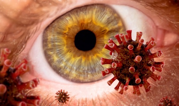
A new medical study has raised serious concerns about the long-term impact of COVID-19 on eye health, especially its potential to cause damage to the retina and optic nerve. Researchers from the Department of Ophthalmology at Recep Tayyip Erdogan University in Rize, Turkey, and the Trabzon Imperial Hospital in Trabzon have found that even after recovery, COVID-19 patients may suffer from hidden neurodegenerative effects in their eyes.
 BREAKING! COVID-19 Found to Trigger Retinal Neurodegeneration
Using advanced imaging technology, the scientists compared the eye health of 78 individuals who had recovered from COVID-19 with 56 healthy individuals of similar age and sex. While no differences were found in basic optic nerve and outer retina measurements, one alarming finding stood out—there was a noticeable thinning of the ganglion cell layer in the retinas of those who had previously contracted COVID-19.
This
Medical News report explores the study’s implications and why even recovered patients might need regular eye checks to catch any silent, progressive damage.
The Ganglion Cell Layer and Its Role in Vision
The ganglion cell layer, located in the retina, is critical for transmitting visual signals from the eye to the brain. Damage to this layer has been linked to several neurodegenerative conditions, such as glaucoma and even Alzheimer's disease. The new findings suggest that COVID-19 might subtly affect this part of the eye, potentially causing long-term or even permanent visual consequences.
Using spectral-domain optical coherence tomography (SD-OCT), a non-invasive imaging tool, the researchers measured the thickness of various eye layers. They found that the retinal nerve fiber layer (RNFL) and the optic nerve head (ONH) parameters remained normal in COVID-19 survivors. However, there was a statistically significant thinning of the ganglion cell–inner plexiform layer (GC-IPL) across all measured sectors in the post-COVID group.
For example, the average GC-IPL thickness was about 7.4% lower in recovered patients than in healthy individuals. While all participants still had normal vision at the time of the study, this difference could signal early-stage degeneration, warranting future monitoring.
Uncovering the Mechanism Behind Retinal Damage
So how does COVID-19 potentially damage the eyes? The researchers believe several mechanisms could be involved. One key factor is the virus’s ability to bind to ACE2 receptors, which are not only present in the lungs but also in the eyes—including the retina, retinal pigment epithelium, and choroid. Once the virus enters these tissues, it may trigger inflammation, disrupt blood vessels, and cause direct cell damage.
Another possible route is through the so-called cytokine storm—a hyperinflammatory response common in severe COVID-19 cases. This excessive inflammation may result in reduced blood fl
ow and oxygen to retinal tissues, causing the thinning observed in the study. The GC-IPL layer is particularly sensitive to such changes, which may explain its selective vulnerability.
Key Study Findings in Detail
-No differences were observed in the optic nerve head (ONH) parameters or retinal nerve fiber layer (RNFL) between the two groups.
-Subfoveal choroidal thickness also remained similar in both recovered and healthy participants.
-Significant thinning was seen in the GC-IPL across all quadrants in recovered COVID-19 patients.
-Despite the thinning, all patients maintained a best-corrected visual acuity of 20/20 and had normal intraocular pressure.
The researchers pointed out that while the ganglion cell layer thinning was relatively mild and within normal population variability, its presence across all quadrants and in multiple subjects signals a pattern that could be linked to COVID-19 exposure.
What This Means for Recovered COVID-19 Patients
Although the study’s participants had no visual complaints and maintained good vision, the changes detected through OCT suggest that COVID-19 could leave subtle but meaningful traces in the eyes. Since the thinning of the GC-IPL can be an early sign of neurodegeneration, the study advocates for more widespread use of OCT imaging in recovered patients, especially those who report ongoing neurological symptoms or visual disturbances.
There’s also a call for more long-term studies to track these patients over time. It’s still unclear whether the thinning observed will progress, remain stable, or eventually resolve. Some earlier studies suggest that these retinal changes may be reversible over months, while others indicate possible permanent effects.
Limitations and the Need for Ongoing Research
The study does have limitations. It was conducted at a single center with a relatively small number of participants. More importantly, it lacked baseline pre-COVID eye scans, which makes it difficult to confirm whether the thinning occurred due to the infection or pre-existed it.
Nonetheless, the research adds to growing evidence that COVID-19 is not just a respiratory illness but a systemic disease that can affect multiple organs, including the eyes. It also reinforces the importance of long-term monitoring and personalized care for those recovering from the virus.
A Cautionary Takeaway
The conclusions from this study serve as a reminder that COVID-19 may leave behind silent damage, even in individuals who appear to have fully recovered. The early and selective thinning of the ganglion cell layer in the retina highlights a possible neurodegenerative process that deserves further investigation.
Regular eye check-ups using non-invasive OCT technology may be advisable for people who have had COVID-19, especially if they notice even minor visual changes. More importantly, these subtle eye changes could also provide insights into broader neurological complications, helping to guide future diagnostic and therapeutic strategies for long COVID.
The study findings were published in the peer reviewed journal: Diagnostics.
https://www.mdpi.com/2075-4418/15/10/1195
For the latest COVID-19 News, keep on logging to Thailand
Medical News.
Read Also:
https://www.thailandmedical.news/news/sars-cov-2-also-targets-the-nervous-system-of-eyes-causing-ocular-neuroinflammation
https://www.thailandmedical.news/news/breaking-even-mild-covid-19-can-cause-microvasculature-changes-of-the-optic-nerve
https://www.thailandmedical.news/news/breaking-covid-19-news-study-claims-that-omicron-infections-can-lead-to-acute-primary-angle-closure-and-also-primary-angle-closure-glaucoma
https://www.thailandmedical.news/articles/coronavirus
https://www.thailandmedical.news/pages/thailand_doctors_listings


 Share
Share
 Tweet
Tweet
 Share
Share