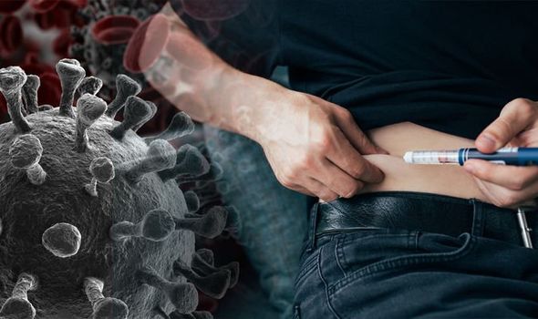
Scientists At Odds On What’s Causing Diabetes In COVID-19 Patients As Two Studies Show That ACE-2 Is Not Expressed In Pancreatic Beta Cells
Source: COVID-19 And Diabetes Sep 02, 2020 5 years, 10 months, 1 week, 2 days, 5 hours, 44 minutes ago
COVID-19 related diabetes being found in SARS-CoV-2 infected
patients who were previously healthy and never had the disease before is causing a dilemma among the medical community as studies are showing that ACE-2 expression is not found in the pancreatic beta cells although it is found in other areas of the pancreas and to date no SARS-CoV-2 virus particles were ever detected in the pancreas even in autopsies.
In the first study by researchers from the Vanderbilt University Medical Center and the University of Pennsylvania Perelman School of Medicine, it was found that both ACE2 and TMPRSS2 that are required for SARS-CoV-2 entry into the human host cell are lacking in the beta cells of the pancreatic islet. These findings rules out the direct injury hypothesis for impaired glucose control in patients with COVID-19 disease.
The study findings for this research were published on a preprint server and have yet to be peer-reviewed.
https://www.biorxiv.org/content/10.1101/2020.08.31.275719v1
The study team first used data from two bulk RNA-sequencing (RNA-seq) datasets, comparing the mRNA expression for these two enzymes in alpha and beta cells from human pancreatic islet tissue with that of important genes encoding, for instance, transcription factors, in islet cells. These genes are typically expressed at low levels in these cells. The median level of ACE2 and TMPRSS2 mRNA expression however was significantly lower than the mRNA for these islet-enriched genes. That is, these two mRNAs were present at 84% and 92% lower expression levels compared to those of some DNA-binding transcription factors within the alpha and beta islet cells.
The team also found that single-cell (sc) RNA-seq analysis of human pancreatic islets demonstrated the absence or minimal expression of mRNAs transcribing the ACE2 and TMPRSS2 genes. The results they used include those obtained from over 25,000 cells of the Human Pancreas Analysis Program (HPAP) database donated by 11 healthy donors, as well as three smaller datasets. Yet, they found ACE2/TMPRSS2 expression in less than 1% of beta cells across all datasets.
Interestingly the exocrine cells showed ACE2 positivity at moderate to high levels for 5% of endothelial cells and pericytes, in the HPAP dataset, but only up to 3% in the other three.
However this difference is not significant and is most likely to be due to varying numbers of cells in each analysis, by up to 20 times more in the HPAP database compared to the smallest set. A pooled analysis shows that overall, less than 1% of acinar or ductal cells had ACE2 expression, but 35% of both kinds of cells expressed TMPRSS2. In contrast, in the smallest study, ACE2 and TMPRSS2 expression in these cells were at the level of 20% and over 75%, respectively.
The study team found that both key enzymes for SARS-CoV-2 infection were expressed together in less than 1% of four cell types (acinar, ductal, endothelial, and stellate) in three datasets. In the fourth, the percentage of co-expression was 5% and 15% for acinar and ductal cells, respectively.
The study te
am points out, “Notably, no islet endocrine cells co-expressed ACE2 and TMPRSS2 in any of these four datasets.”
The team moved from transcriptomics to direct visualization. They used immunostaining on islet cryosections with the same ACE2 antibody reported by earlier investigators. However, they failed to detect ACE2 on beta cells, but only on the microvasculature.
The study team points out that the difference might possibly be due to the use of stem cell-derived beta cells, which are considered to be juvenile-like cells rather than functionally mature, or the immortalized beta cells in culture, in an earlier study.
In order to rule out this possibility, the team then repeated the process utilizing sections from pancreatic tissue obtained from children aged 5 years or less (one was only 5 days old). However, they still did not find ACE2.
The researchers also looked at donor pancreatic tissue from diabetic and non-diabetic donors. The diabetic donors had either type 1 or type 2 diabetes. Here again, ACE2 was not found on either alpha or beta cells, but the microvascular structures in both the healthy donors and those with type 2 diabetes.
The team repeated their analysis with three other ACE2 antibodies used by several other researchers, but failed to find this protein on islet cells.
The study team also did not find TMPRSS2 within islets in healthy or type 1 diabetic donors, but it was localized on exocrine tissue, on duct epithelium.
The team concluded that these proteins are not found on islet endocrine cells from either healthy or diabetic donors. The scientists now think that the earlier findings may be due to key differences in the way the experiment was set up and perhaps artifacts.
Based on a recent silico study that indicated the possibility of viral binding to human dipeptidyl peptidase 4 (DPP4) in enabling viral entry, they searched for this protein on the human pancreas. They found that it was localized to alpha but not beta cells of all donors, diabetic or not.
https://www.frontiersin.org/articles/10.3389/fphar.2020.01161/full
The study findings agree with the transcriptomic analysis based on all four datasets, which also demonstrated that these cells were DPP4-enriched. However, the absence of TMPRSS2 indicates, they say, that DPP4 is probably not related to viral cell entry into beta cells.
The team also looked into the staining pattern of ACE2 in the non-endocrine cells of the pancreas, specifically to find out if it was localized in the microvasculature. They concluded that this was the case, with the perivascular capillaries of the islets as well as exocrine tissue showing ACE2 expression.
The team suggest that this protein is expressed on pericytes, the cells that envelope capillary endothelial cells. However, since there is no TMPRSS2, this cell would not be targeted by SARS-CoV-2. And in fact, though both these proteins are found on duct epithelium, they are not found together.
.jpg) ACE2 and TMPRSS2 Protein is Not Detected by Immunofluorescence in a or b Cells from Normal, T2D or T1D adult donors. SARS-CoV-2 cell entry markers ACE2 (antibody ab15348) and TMPRSS2, both shown in red, are not detected in islet a cells (GCG, blue) or b cells (INS, green) in pancreatic sections from adult donors without diabetes (A-H) or donors with type 2 (I-N) or type 1 (O-V) diabetes. Insets are depicted by a yellow box. DAPI (white). Scale bars are 100 μm (A-V) and 25 μm (Insets). Human islet and pancreatic donor information is available in Table S1 (A-D, donors N3, N7, N9, N8; E-H, donors N14, N12, N11, N10; I-L, donors 2L, 2B, 2G, 2I; M-N, donors 2H, 2G; O-R, donors 1B, 1D, 1C, 1A; S-V, donors 1H, 1K, 1J, 1G).
ACE2 and TMPRSS2 Protein is Not Detected by Immunofluorescence in a or b Cells from Normal, T2D or T1D adult donors. SARS-CoV-2 cell entry markers ACE2 (antibody ab15348) and TMPRSS2, both shown in red, are not detected in islet a cells (GCG, blue) or b cells (INS, green) in pancreatic sections from adult donors without diabetes (A-H) or donors with type 2 (I-N) or type 1 (O-V) diabetes. Insets are depicted by a yellow box. DAPI (white). Scale bars are 100 μm (A-V) and 25 μm (Insets). Human islet and pancreatic donor information is available in Table S1 (A-D, donors N3, N7, N9, N8; E-H, donors N14, N12, N11, N10; I-L, donors 2L, 2B, 2G, 2I; M-N, donors 2H, 2G; O-R, donors 1B, 1D, 1C, 1A; S-V, donors 1H, 1K, 1J, 1G).
Interestingly TMPRSS2 is on the apical surface of duct cells in the exocrine pancreas, but not ACE2. In the few instances where they are both found together, they are separately localized. The researchers say they may represent a possible viral target.
Certain limitations of the current study include the fact that the researchers relied on the demonstration of these proteins and not on looking at viral binding or entry into the beta cells. Finally, they did not measure the expression of these proteins in diabetic patients with COVID-19, so that they cannot rule out changes in these receptors in such patients.
The researchers concluded “Altogether, these findings greatly reduce the likelihood that SARS-CoV2 can bind and enter human b cells and have direct cytotoxicity.” Instead, they favor the explanation that glucose homeostasis is probably impacted by inflammation or damage to liver, muscle, or fat cells, which needs to be borne out by further research.
In the second study by researchers from the University of Florida, Indiana University, Icahn School of Medicine at Mount Sinai, Louisiana State University, Baylor College of Medicine and the University of Miami, the team focused on the effect of SARS-CoV-2 infection on β-cells on type 1/type 2 diabetes mellitus (T1D or T2D). They examined ACE2 expression in both exocrine and endocrine cells of the pancreas, but particularly the islet of Langerhans, which houses the β-cells.
They too found no evidence of SARS-CoV-2 particles or ACE2 receptors in the pancreatic beta cells. The study findings were also published on a preprint server and have yet to be peer-reviewed.
https://www.biorxiv.org/content/10.1101/2020.08.31.270736v1
Utilizing publicly available single-cell RNA sequencing (scRNAseq) data, along with fluorescence in situ hybridization (FISH), immunohistochemistry (IHC), and immunofluorescence (IF) studies to directly observe gene and protein expression of ACE2, they looked for this protein in human tissue using four commercially available ACE2 antibodies with IHC/immunoblot controls. They also evaluated the expression of the viral nucleocapsid (N) protein in pancreatic tissues from deceased COVID-19 patients derived from post-mortem studies.
The study team also examined tissue from 36 COVID-19-negative donors who did not have diabetes, aged 0-72 years. They found that the ACE2 staining percentage of tissue increases steadily from birth to a peak in the adolescent years, to plateau in early adult life and finally decline over the age of 50 years.
Significantly at all ages, ACE2 is expressed in pancreatic duct epithelial cells and microvasculature within the endocrine regions, but not on α-cells or β-cells. These findings were confirmed using scRNAseq and smFISH gene expression data.
The study team also looked simultaneously at scRNAseq data from five datasets, including 22 non-diabetic and 8 type 2 diabetics, finding low ACE2 expression in most islet cell subsets. This showed that normal pancreas from donors without COVID-19 expresses ACE2 mainly in duct cells and endothelium of the small blood vessels.
It was also observed that for the donor samples ie in patients without diabetes, less than 2% of cells in the pancreas expressed ACE2, at 4% of acinar cells and over 5% of ductal cells. Type 2 diabetics showed ACE2 expression in 8% of both acinar and ductal cells. ACE2 expression in the islet cells was thus comparably low between diabetic and non-diabetic donors, while TMPRSS2 expression was above 50% in acinar and ductal cells. However, the expression of TMPRSS2 was low in most endocrine cells in the pancreas.
.jpg) Pathological changes in pancreata of COVID-19 patients. (A) Pancreas tissue section from COVID-19 Patient 1 stained for H&E. Inset highlights fibrotic center with residual acinar cells and islet surrounding ductules. Scale bars: 3mm, inset 200µm. (B) Pancreas tissue section from COVID-19 Patient 2 stained for H&E. Inset highlights microthrombus without adjacent hemorrhages. Scale bars: 4mm, inset 400µm. (C) Pancreas tissue section of COVID-19 Patient 3 stained for H&E. Inset highlights a large, irregularly shaped pancreatic islet surrounded by fibrotic tissue. Scale bars: 4 mm, inset 200µm. (D) Representative pancreas tissue sections from three COVID-19 patients stained for ACE2, insulin (INS) and glucagon (GCG). Scale bars: 200µm. (E) SARS-CoV-2 NP observed in intralobular ducts (d) near an islet in the pancreas of COVID19 Patient 1. Scale bars: 10µm. (F) Representative image of multiple ducts showing SARS-CoV-2 NP positivity in the pancreas of COVID-19 Patient 1. Scale bar: 20µm.
Pathological changes in pancreata of COVID-19 patients. (A) Pancreas tissue section from COVID-19 Patient 1 stained for H&E. Inset highlights fibrotic center with residual acinar cells and islet surrounding ductules. Scale bars: 3mm, inset 200µm. (B) Pancreas tissue section from COVID-19 Patient 2 stained for H&E. Inset highlights microthrombus without adjacent hemorrhages. Scale bars: 4mm, inset 400µm. (C) Pancreas tissue section of COVID-19 Patient 3 stained for H&E. Inset highlights a large, irregularly shaped pancreatic islet surrounded by fibrotic tissue. Scale bars: 4 mm, inset 200µm. (D) Representative pancreas tissue sections from three COVID-19 patients stained for ACE2, insulin (INS) and glucagon (GCG). Scale bars: 200µm. (E) SARS-CoV-2 NP observed in intralobular ducts (d) near an islet in the pancreas of COVID19 Patient 1. Scale bars: 10µm. (F) Representative image of multiple ducts showing SARS-CoV-2 NP positivity in the pancreas of COVID-19 Patient 1. Scale bar: 20µm.
The team found that both ACE2 and the cell protease TMPRSS2 were expressed at low levels in human pancreatic cells and the islet’s β-cells. These findings were confirmed using direct visualization methods.
The study team also looked at pancreatic tissue retrieved by autopsy of three patients with fatal COVID-19, aged 45-72 years. Two of these patients were known to have type 2 diabetes. They found that in the non-diabetic patient, fatty replacement of the acinar cells, with islets within the fibrotic regions. The other patients showed moderate to numerous islets.
The team commented, “These histopathological findings were compatible with the normal range of expected lesions within the exocrine compartment in pancreata from aged patients and those with Type 2 diabetes.”
Furthermore direct visualization by IHC showed moderate ACE2 expression in duct epithelium and no evidence of viral N protein in the endocrine pancreatic tissue. Instead, N protein was localized to the duct epithelium, widely scattered through the exocrine pancreas, and there were several thrombotic lesions.
The study findings seems to rule out direct viral infection of endocrine β-cells of the pancreas as the reason for the clusters of new diabetes cases or the increased mortality in people with diabetes with COVID-19. Indeed, the duct epithelium and microvascular endothelium seem to be more likely viral targets.
Both studies are causing a dilemma into understanding what is triggering the diabetes in otherwise health COVID-19 patients.
https://www.thailandmedical.news/news/breaking-medical-experts-warn-that-covid-19-could-trigger-diabetes-in-otherwise-healthy-individuals-who-never-had-the-condition-before-
https://www.thailandmedical.news/news/breaking-covid-19-and-children-uk-researchers-warn-that-covid-19-could-be-linked-to-onset-of-type-one-diabetes-and-diabetic-ketoacidosis-in-children
In earlier studies it was found that many COVID-19 patients were developing diabetes and due to the occurrence of diabetic ketoacidosis and hyperosmolar hyperglycemia, it led to the proposition that SARS-CoV-2 is a diabetogenic virus, by causing toxicity to the beta-cells of the pancreatic islets that secrete insulin.
https://www.researchgate.net/publication/24245824_Binding_of_SARS_coronavirus_to_its_receptor_damages_islets_and_causes_acute_diabetes and
https://www.medrxiv.org/content/10.1101/2020.02.28.20029181v1.full.pdf
The earlier coronavirus, SARS-CoV, which caused a more limited outbreak between 2002 and 2004, also uses the ACE2 receptor. Prior research on autopsy samples from patients who died of this infection reported that ACE2 was found on pancreatic islet cells from one donor (though the type of cell was not clear). Thus, they first floated the idea that virus-ACE2 engagement causes islet damage and triggers acute diabetes. This was reversible once the infection was cleared.
https://www.ncbi.nlm.nih.gov/pmc/articles/PMC7088164/
A research reported recently that they induced beta-like cells from human pluripotent stem cells (hPSCs). Tests on these cells, as well as the beta cells from human pancreatic islets isolated from the organ, were found to express ACE2, supporting their hypothesis of direct viral injury to these cells.
https://www.sciencedirect.com/science/article/pii/S1934590920302824?via%3Dihub
The fact does remain that SARS-CoV-2 coronavirus in COVID-19 patients that were otherwise healthy and never had the diabetes before but how that is being caused remains to be addressed and further studies are needed.
For more on
COVID-19 and diabetes, keep on logging to Thailand Medical News.


