Swedish Study Shows SARS-COV-2 Also Causes Myelin Oligodendrocyte Glycoprotein-Associated Disorder (MOGAD), A CNS Autoimmune Disease!
NeuroCOVID: A new study by researchers from the University of Gothenburg-Sweden and Sahlgrenska University Hospital-Sweden involving four clinical case reports has found that SARS-CoV-2 ca also trigger a CNS autoimmune disease called Myelin Oligodendrocyte Glycoprotein-Associated Disorder or MOGAD.
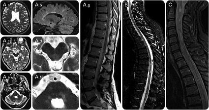 MRI Features Of Individuals With Myelin oligodendrocyte glycoprotein antibody disorders (MOGAD)
Myelin oligodendrocyte glycoprotein antibody disorders (MOGAD)
MRI Features Of Individuals With Myelin oligodendrocyte glycoprotein antibody disorders (MOGAD)
Myelin oligodendrocyte glycoprotein antibody disorders (MOGAD) is an idiopathic, inflammatory, demyelinating disease of the central nervous system (CNS). MOG is a glycoprotein uniquely expressed in oligodendrocytes in the CNS. MOG antibodies were originally thought to be involved in multiple sclerosis (MS), but subsequent studies found it to be a distinct disease.
MOGAD have many similarities to neuromyelitis optica but several studies have demonstrated they have unique clinical features, treatment response, and prognosis. MOGAD have several clinical presentations. One of the most common presentations is similar to neuromyelitis spectrum disorder (NMOSD) with recurrent optic neuritis, transverse myelitis, or both. Another common presentation is that of acute disseminated encephalomyelitis (ADEM). ADEM can present with typical symptoms of encephalitis including decreased consciousness, headache, and behavioral changes. Seizures can also occur in the context of MOGAD associated ADEM presentations and occur at varying rates. Other presentations include brainstem syndrome and short segment transverse myelitis.
MOG antibody disease preferentially causes inflammation in the optic nerve, but can also cause inflammation in the spinal cord, brain, and brainstem.
Typical symptoms can include:
-Loss or blurring of vision in one or both eyes
-Loss of color vision
-Paralysis (no motor function) of a limb or limbs
-Paraparesis (weakness) of a limb or limbs
-Loss of sensation
-Loss of bladder or bowel control
-Profound bladder retention
-Seizures
The actual and detailed cause of MOGAD is unknown.
It has already been known that SARS-COV-2 infections frequently cause neurological disorders and is sometimes associated with onset of autoimmune diseases affecting the nervous system.
There have been reports of single cases of MOGAD in patients with COVID-19 infection.
The
NeuroCOVID study team reports a series of SARS-CoV-2 positive patients that developed MOGAD, but a homology search did not support a cross-reactive immune response to SARS-CoV-2 spike-protein and MOG.
The case reports and study findings were published in the peer reviewed journal: Annals of Clinical and Translational Neurology. (A publication of the American Neurological Association.)
https://onlinelibrary.wiley.com/doi/10.1002/acn3.51609
According to the study team, the COVID-19 disease precipitate
s neurological symptoms for which the triggering of autoantibody diseases in the host could be a causative factor.
MOGAD or Myelin Oligodendrocyte Glycoprotein-Associated Disorder - a demyelinating condition of the central nervous system (CNS), could be monophasic or relapsing and is characterized by the presence of antibodies to the myelin oligodendrocyte glycoprotein (MOG).
Numerous reports suggest that severe acute respiratory syndrome coronavirus 2 (SARS-CoV-2) may induce MOGAD, owing to the higher occurrence of SARS-CoV-2 immunoglobulin (Ig)G antibodies in these patients. Another study depicted that anti-MOG positivity was shown by just one among 15 post-COVID-19 patients with acute disseminated encephalomyelitis (ADEM) or acute hemorrhagic leukoencephalitis.
These case studies also reviewed previous case reports with similar associations - emphasizing the role of SARS-CoV-2 in the pathogenesis of MOGAD. All four patients were diagnosed with MOGAD post-COVID-19 in Västra Götaland County.
Case Study One - Acute Disseminated Encephalomyelitis
This cases study involves a 25-year-old lady presented with fever and headache and was detected PCR-positive for SARS-CoV-2. She experienced severe paraparesis, hypesthesia, decreased consciousness, as well as urinary retention for two weeks.
MRI or Magnetic resonance imaging of the brain and spinal cord showed multiple non-enhancing cerebral parenchymal and spinal cervicothoracic high-intensity lesions (T2). The cerebrospinal fluid (CSF) showed two oligoclonal IgG bands.
The female patient was diagnosed with ADEM; she was started on i.v. methylprednisolone 1 g for 5 days. The diagnostic workup for SARS-CoV-2 and serum IgG aquaporin-4 (AQP4)-antibodies were negative while anti-MOG IgG showed a positive result (1:1000).
The same MRI was repeated after two weeks, which exhibited new non-contrast enhancing high signal lesions on T2/fluid-attenuated inversion recovery (FLAIR). These lesions were observed in the corpus callosum, pons, mesencephalon, and progress of intramedullary lesions in the cervical and thoracic spinal cord, and included contrast enhancement from (thoracic spines) Th6-Th9.
The infected individual was prescribed a new course of intravenous steroids, which was followed by plasmapheresis sessions for five days. Thereafter, methotrexate and slow tapering of oral prednisolone was given.
It was found that after four weeks, the patient elicited a relapse of optic neuritis with edema of the optic disc. MRI showed a new lesion in the cerebellar peduncle. The patient showed complete radiological (MRI) resolution of the cerebral and spinal lesions four months after MOGAD onset after undergoing a new course of high dose i.v. steroids.
The female patient has continued on prednisolone 10 mg daily and methotrexate 15 mg weekly. She had a minor lower extremity dysesthesia and bladder dysfunction.
Case Study Two - Bilateral Optic Neuritis And Myelitis
The second cases study involves a 20-year-old male who was PCR positive for SARS-CoV-2 with minor respiratory symptoms. He had a history of personality disorder and substance abuse. The patient presented with a gradual onset of headache, back pain, urinary retention, reduced vision, photo-and phonophobia, severe paraparesis, and hemianesthesia, eight weeks post-infection.
A detailed neuro-ophthalmologic examination and OCT showed bilateral optic disc edema, impaired visual acuity and color vision indicating optic neuritis.
A detailed MRI of the spinal cord exhibited conspicuous medullary T2 lesions from Th9 to conus medullaris, along with small focal lesions at Th7 and (cervical spine) C6 levels, and a slight contrast enhancement at Th9-Th11. Serum MOG-antibody test showed positive (1:100 titer), whereas the anti-AQP4 antibody test was negative.
The male patient was negative for infectious or rheumatological findings. He was prescribed plasmapheresis (five consecutive days), followed by i.v. methylprednisolone 1 g/day for 3 days. Thereafter, the patient commenced on oral prednisolone tapered to 10 mg/day and 15 mg methotrexate weekly. Six months of therapy led to improvements and he had only moderate residual paraparesis.
Case Study Three - Bilateral Optic Neuritis
The third cases involved a 29-year-old man presented with bilateral visual impairment and frontal headache. He had been diagnosed with COVID-19 – 15 days prior. No other relevant medical or drug history was evident.
The patient was diagnosed with papillitis due to optic neuritis; OCT revealed bilateral optic disc edema. A thorough neurological examination did not elicit any deficit; the MRI brain was normal. Lumbar puncture depicted normal opening pressure, CSF white blood cells, and protein levels, while the oligoclonal IgG bands were negative.
This male patient was started on methylprednisolone i.v. 1 g/day for 3 days. The patient was then prescribed oral prednisolone tapered at 10 mg/day.
The serum AQP4-antibodies were negative and anti-MOG IgG was positive (1:100 titer). One month after post-therapy, the patient’s vision normalized. Follow-up after six weeks rendered an anti-MOG serum titer of 1:10 which became negative four months after the optic neuritis diagnosis.
Case Study Four - Unilateral Optic Neuritis
The last cases involved a 60-year-old Chinese woman had COVID-19 symptoms of fever, cold, dyspnea, and myalgia. She was PCR positive for SARS-CoV-2. She was admitted to the intensive care unit (ICU) due to respiratory insufficiency and was put on oxygen; the patient was also administered oral betamethasone and rivaroxaban. After improvements in her symptoms, the patient was discharged.
However, fifty-four days after the onset of COVID-19 symptoms the lady complained of right-sided eye pain and was diagnosed with unilateral optic neuritis with papillitis. On examination, she had reduced visual acuity ie 20/100.
A detailed MRI displayed edema and contrast enhancement in the right optic nerve without a significant pathology in the cerebral parenchyma or the spinal cord. The CSF cell count, albumin, and neurofilament light chain (NfL) were normal with no oligoclonal IgG bands.
She was given methylprednisolone 1 g/day for 3 days and then, slow tapering prednisolone. Serology testing disclosed a negative anti-AQP4 and positive MOG-antibodies (1:1000 titer).
Conclusions Drawn From The 4 Case Studies
The study team reported four MOGAD diagnoses shortly after contracting COVID-19. The evidence was consistent with the post-infectious immune-mediated reaction associated with SARS-CoV-2 infection. The mean latency period after COVID-19 onset to MOGAD manifestation was 33 days––with a range of 14-56 days. A similar latency of 10-42 days was observed in patients with SARS-CoV-2 infection who presented with acute transverse myelitis as a post-infectious immune-mediated reaction.
The study findings show that MOGAD onset was closely associated with prior COVID-19 and SARS-CoV-2 infection, and this was implicated in the higher MOGAD incidence in Västra Götaland County during the pandemic.
For more on
Myelin Oligodendrocyte Glycoprotein Antibody Disorders (MOGAD) And SARS-CoV-2, keep on logging in to Thailand
Medical News.


 Share
Share
 Tweet
Tweet
 Share
Share


