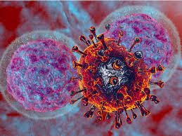
Yale Study Discovers That SARS-CoV-2 Proteins ORF7a And ORF3a Downregulates MHC-I Expression Aiding In Immune Evasion And Immune Dysfunction!
Source: Medical News - COVID-19 Immunology May 24, 2022 3 years, 8 months, 3 weeks, 1 day, 23 hours, 50 minutes ago
COVID-19 Immunology: A new study by researchers from Yale University School of Medicine-USA has found that SARS-CoV-2 accessory proteins ORF7a and ORF3a downregulates MHC-I expression aiding in immune evasion and possible immune dysfunction!

MHC class I molecules (MHC-I) are cell surface recognition elements expressed on virtually all somatic cells. These molecules sample peptides generated within the cell and signal the cell's physiological state to effector cells of the immune system, both T lymphocytes and natural killer (NK) cells.
MHC class I molecules are one of two primary classes of major histocompatibility complex (MHC) molecules (the other being MHC class II) and they also occur on platelets, but not on red blood cells. Their function is to display peptide fragments of proteins from within the cell to cytotoxic T cells; this will trigger an immediate response from the immune system against a particular non-self antigen displayed with the help of an MHC class I protein. Because MHC class I molecules present peptides derived from cytosolic proteins, the pathway of MHC class I presentation is often called cytosolic or endogenous pathway
The major histocompatibility complex class I (MHC-I) molecules, which are dimers of a glycosylated polymorphic transmembrane heavy chain and the small protein β2-microglobulin (β2m), bind peptides in the endoplasmic reticulum that are generated by the cytosolic turnover of cellular proteins.
In virus-infected cells these peptides may include those derived from viral proteins. Peptide-MHC-I complexes then traffic through the secretory pathway and are displayed at the cell surface where those containing viral peptides can be detected by CD8+ T lymphocytes that kill infected cells.
Most viruses enhance their in vivo survival by encoding genes that downregulate MHC-I expression to avoid CD8+ T cell recognition.
The
COVID-19 Immunology study findings report that two accessory proteins encoded by SARS-CoV-2, the pathogenic agent of the ongoing COVID-19 pandemic, downregulate MHC-I expression using distinct mechanisms.
The first protein, ORF3a, a viroporin, reduces global trafficking of proteins, including MHC-I, through the secretory pathway.
The other, ORF7a, interacts specifically with the MHC-I heavy chain, acting as a molecular mimic of β2m to inhibit its association. This slows the exit of properly assembled MHC-I molecules from the endoplasmic reticulum.
The study team demonstrated that ORF7a reduces antigen presentation by the human MHC-I allele HLA-A*02:01. Thus, both ORF3a and ORF7a act post-translationally in the secretory pathway to lower surface MHC-I expression, with ORF7a exhibiting a novel and specific mechanism that allows immune evasion by SARS-CoV-2.
Importantly, it should be noted that viruses may down-regulate MHC class I expression on infected cells to avoid elimination by cytotoxic T cells.
The study findings show that the accessory proteins ORF7a and ORF3a of SARS-CoV-2 mediate this function and delineate the two distinct mechanisms involved. While ORF3a inhibits global protein trafficking to the cell surface, ORF7a acts specifically on MHC-I by competin
g with β2m for binding to the MHC-I heavy chain. This is the first account of molecular mimicry of β2m as a viral mechanism of MHC-I down-regulation to facilitate immune evasion.
The study findings were published on a preprint server and is currently being peer reviewed.
https://www.biorxiv.org/content/10.1101/2022.05.17.492198v1
MHC-I is a heterodimer of beta-2-microglobulin (β2m) and a glycosylated heavy chain. The MHC-I molecules are loaded with peptides in the endoplasmic reticulum (ER), including viral peptides. MHC-I-peptide complexes are trafficked to the cell surface where self, non-self, or viral peptides are displayed and presented to the immune cells. The cluster of differentiation 8 (CD8+) T cells identify and kill cells with non-self-peptides.
Typically, it has been found that viruses downregulate MHC-I expression to prevent elimination by the adaptive arm of the immune system. This could be achieved by downregulating transcription of MHC-I genes or inhibition of MHC-I function. It has been reported that SARS-CoV-2 infection downregulates a specific MHC-I allele, HLA-A*02:01 (HLA-A2), with open reading frame-6 (ORF6) and ORF8 implicated in the process.
The study team investigated the roles of ORF3a and ORF7a proteins of SARS-CoV-2 because these have been predicted to localize to the secretory pathway.
For the study, Vero E6 or HEK293T-hACE2 cells expressing angiotensin-converting enzyme 2 (ACE2) were infected with SARS-CoV-2 WA1 at 10 multiplicity of infection (MOI) for 24 hours. When MHC-I expression was estimated using flow cytometry, a 20% – 30% decline in surface expression was noted in the infected cells relative to controls (non-infected cells).
The study team then separately expressed ORF3a, ORF7a, and ORF8 in HEK293T or HeLaM cells, and after 24 hours of transfection, the study team noted a 25% to 30% reduction in surface expression of MHC-I.
Corresponding author, Dr Peter Cresswell, a Professor from the Department of Immunobiology, Yale University School of Medicine told Thailand
Medical News, “It was also found that the epidermal growth factor receptor (EGFR) and CD47 levels were unaffected by ORF8 or ORF7a, whereas ORF3a downregulated CD47 and EGFR in both cell lines.”
Importantly, this implied that ORF7a or ORF8 expression affects MHC-I levels, while ORF3a expression impairs protein trafficking via the secretory pathway.
The ORF3a protein from SARS-CoV-1 and SARS-CoV-2 were individually expressed in HeLaM cells, and Golgi morphology was examined by confocal microscopy. The study team detected Golgi fragmentation in both cases, while non-infected cells had intact Golgi system. A transient ORF3a expression for 24 hours reduced MHC-I levels by approximately 30%.
The study team posits that ORF3a downregulates MHC-I on the surface by disrupting the Golgi apparatus and blocking protein trafficking to the cell surface.
In further experiments involving inducible expression of ORF7a on doxycycline (Dox) treatment in HeLaM cells (henceforth HeLaM-iORF7a cells), the study team observed that MHC-I expression on the surface was 20% lower, albeit the messenger ribonucleic acid levels (mRNA) levels remained unchanged relative to controls. This confirmed that the downregulation of MHC-I was not transcriptional.
The MHC-I levels in ORF7a-expressing HeLaM cells were similar to controls in lysates. Hence, the study team investigated whether ORF7a affected intracellular MHC-I distribution and found that the MHC-I export rate from the ER was slowed by ORF7a expression.
The study team next observed co-immunoprecipitation of ORF7a with MHC-I heavy chain but not β2m, or MHC-I-peptide complex, indicating that ORF7a interferes with β2m association with the MHC-I heavy chain.
A β2m-encoding plasmid or a control plasmid (no β2m) was transiently expressed in HeLaM-iORF7a cells. Expectedly, cells with a control plasmid showed reduced MHC-I surface expression upon Dox treatment relative to untreated cells (no Dox). However, this was eliminated when β2m was expressed, implying that excess β2m levels could overcome ORF7a action. Confocal examination revealed localization of ORF7a to both the Golgi system and ER.
The study team also generated a mutant variant of ORF7a (ORF7a-ARA) wherein lysine residues from a dibasic sequence (an ER-retrieval sequence) at the C-terminus were substituted with alanine residues.
The study team observed that this mutant was primarily localized to the Golgi system. In the presence of ORF7a, MHC-I was retained to ER, while this was not the case with ORF7a-ARA.
The team also generated Dox-inducible versions of ORF7a or ORF7a-ARA in HEK293T-hACE2 cells to investigate the effects on surface levels of MHC-I and antigen presentation by HLA-A2 (since HeLaM cells lack this allele).
Interestingly, surface MHC-I levels were reduced by 40% when ORF7a expression was induced, but not with ORF7a-ARA induction.
The study team last used a specific MHC-I-peptide complex, with latent membrane protein 2A (LMP2A) from Epstein-Barr virus as the peptide. LMP2A is presented by and binds to HLA-A2 with high affinity.
To this end, HEK293T-hACE2 cells and their ORF7a or ORF7a-ARA inducible versions were transduced with human influenza hemagglutinin (HA)-tagged fusion construct of ubiquitin: LMP2A-derived peptide (pLMP2A).The presentation of the LMP2A-HLA-A2 complex was assessed using a specific monoclonal antibody. pLMP2A and ORF7a were expressed upon treatment with Dox, but only in HA-positive cells with ORF7a but not ORF7a-ARA, the presentation of pLMP2A was reduced due to decreased HLA-A2 levels mediated by ORF7a.
The research findings study demonstrated that the two SARS-CoV-2 accessory proteins (ORF3a and ORF7a) downregulate MHC-I with distinct mechanisms. MHC-I downregulation by ORF3a was more general with the inhibition of protein trafficking, whereas, by ORF7a, it was specific in that it interacts with the MHC-I heavy chain, slowing the export of MHC-I-peptide complexes.
It was noted that the downregulation of MHC-I by ORF7a was attenuated when the ER retrieval sequence was mutated by replacing lysine with alanine.
The study findings conclude that ORF3a and ORF7a reduce surface MHC-I levels, with the downregulation mediated by ORF7a being more specific and novel, allowing immune evasion by SARS-CoV-2.
Researchers are also worried that this study findings also shows one of the many ways that the SARS-CoV-2 virus causes a dysfunctional immune system that could help accelerate secondary opportunistic pathogenic infections in those who were infected with the SARS0CoV-2 virus.
For more on
COVID-19 Immunology, keep on logging to Thailand Medical News.
