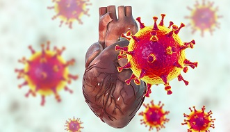
BREAKING! University of Maryland Study Shockingly Finds That SARS-CoV-2 Nsp6 Proteins Can Induce Morphological And Functional Defects In The Heart!
Source: CardioCOVID Jun 07, 2022 3 years, 8 months, 1 week, 2 days, 2 hours, 46 minutes ago
CardioCOVID: A new study by researchers from University of Maryland School of Medicine-USA has found that the SARS-CoV-2 protein Nsp6 is able to induce cardiac defects in the human host through MGA/MAX complex-mediated increased glycolysis! This shocking revelation showed that the Nsp6 proteins can induce morphological as well as functional defects that can eventually lead to heart failures!

Importantly the
CardioCOVID study also found that using the pharmaceutical compound 2-deoxy-D-glucose (2DG) could help prevent COVID-19 induced heart failures!
Yet another interesting finding from the study was that ketogenic diets might also help in better clinical outcomes and also prevent heart failures in COVID-19 infected and Post COVID individuals.
Furthermore, considering the kind of cardiac damage that the various SARS-CoV-2 proteins can cause both directly and indirectly to the human host heart during infection and Post-COVID, drugs such as fluvoxamine which can induce arrythmia should never be used to treat COVID-19. Currently many stupid and garbage doctors are promoting the use of fluvoxamine to treat COVID-19 including claims that it prevents or resolves disease severity.(It should be noted that fluvoxamine has no antiviral effects against the current SARS-CoV-2 variants except exhibiting certain anti-inflammatory properties which again are no longer relevant in the context of the new variants that exhibit a different pathogenesis!) Family members of patients who have died or suffered heart issues while being treated with fluvoxamine to treat COVID-19 should sue the doctors, hospitals and also the individuals, media and social media postings that promote its use.
https://www.thailandmedical.news/news/most-who-have-been-exposed-to-the-proteins-of-the-sars-cov-2-virus-will-have-shortened-lifespans-stop-using-fluvoxamine-for-ba-2-infections
Already COVID-19 presents with a wide variety of symptoms that often include cardiac pathology. The detection of SARS-CoV-2 in cardiac tissue suggests the cardiac pathology is caused by direct virus action, rather than secondary complications.
https://pubmed.ncbi.nlm.nih.gov/32529795/
https://pubmed.ncbi.nlm.nih.gov/32730555/
https://pubmed.ncbi.nlm.nih.gov/32275347/
https://pubmed.ncbi.nlm.nih.gov/34282465/
https://www.ncbi.nlm.nih.gov/pmc/articles/PMC8104461/
https://pubmed.ncbi.nlm.nih.gov/34647998/
https://diagnosticpathology.biomedcentral.com/articles/10.1186/s13000-022-01207-6
gt;
Importantly, elevated levels of cardiac biomarkers-such as, troponins, myoglobin, C-reactive protein, interleukins and natriuretic peptides, all indicative of myocardial injury, have been observed in COVID-19.
https://pubmed.ncbi.nlm.nih.gov/34258904/
The presence of these elevated cardiac biomarkers combined with abnormal echocardiograms that reflect functional deficits, has been associated with a poorer prognosis of disease progression.
https://pubmed.ncbi.nlm.nih.gov/32556199/
https://pubmed.ncbi.nlm.nih.gov/32219356/
https://pubmed.ncbi.nlm.nih.gov/32211816/
To date, cardiovascular complications reported in COVID-19 patients include necrosis, ventricular dysfunction heart failure, and arrhythmia.
https://pubmed.ncbi.nlm.nih.gov/34258904/
There is also evidence of myocardial inflammation (myocarditis) following SARS-CoV-2 69 infection, including in asymptomatic individuals and, the long-term (1-year) risk of cardiovascular disease is considerable in COVID-19 patients, independent of hospitalization.
https://pubmed.ncbi.nlm.nih.gov/32730619/
https://pubmed.ncbi.nlm.nih.gov/32915194/
https://www.nature.com/articles/s41591-022-01689-3
Importantly, the key to treating the SARS-CoV-2 induced cardiovascular pathology likely lies in understanding and targeting the individual virus-host interactions.
https://cellandbioscience.biomedcentral.com/articles/10.1186/s13578-021-00621-5
https://pubmed.ncbi.nlm.nih.gov/34124934/
Many of these have been investigated in large-scale network studies which revealed distinct proteome interaction networks, for many of these interactions specific pharmacological compounds exist.
https://pubmed.ncbi.nlm.nih.gov/32353859/
https://pubmed.ncbi.nlm.nih.gov/33060197/
https://pubmed.ncbi.nlm.nih.gov/32244779/
https://pubmed.ncbi.nlm.nih.gov/33339864/
https://pubmed.ncbi.nlm.nih.gov/33589624/
https://pubmed.ncbi.nlm.nih.gov/33837377/
Studies have recently been published that delve deeper into the individual SARS-CoV-2 proteins, their host interactions and underlying pathomechanisms. These have already revealed that some virus proteins known for their role in virus replication processes, such as SARS-CoV-2 Nsp3 and Nsp5 which encode the (PLpro) and (3CL pro; main protease, Mpro), are also responsible for disrupting host immune signaling processes through specific protein-protein interactions.
https://pubmed.ncbi.nlm.nih.gov/32726803/
https://pubmed.ncbi.nlm.nih.gov/33372854/
These findings suggest that therapeutics targeting these virus proteins specifically could both reduce virus replication and diminish their role in the damaging host pathogenic effects.
The study team from University of Maryland by expressing individual SARS-CoV-2 proteins in the Drosophila heart, demonstrated interaction of virus Nsp6 with host proteins of the MGA/MAX complex (MGA, PCGF6 and TFDP1).
It should be noted that the metabolic pathways that regulate heart development and function, including glycolysis, are highly conserved from fly to human
https://journals.plos.org/plosone/article/comment?id=10.1371/annotation/c7259943-7050-4771-8f79-df744d30dd8f.
Complementing transcriptomic data from the fly heart revealed that this interaction blocks the antagonistic MGA/MAX complex, which shifts the balance towards MYC/MAX and activates glycolysis, with similar findings in mouse cardiomyocytes. Further, the Nsp6-induced glycolysis disrupted cardiac mitochondrial function, known to increase reactive oxygen species (ROS) in heart failure; this could explain COVID-19-associated cardiac pathology.
It was found that inhibiting the glycolysis pathway by 2-deoxy-D-glucose (2DG) treatment attenuated the Nsp6-induced cardiac phenotype in fly and mice; thus, suggesting glycolysis as a potential pharmacological target for treating COVID-19-associated heart failure.
The study findings were published on a preprint server and are currently being peer reviewed for publication into the Nature journal.
https://www.researchsquare.com/article/rs-1677754/v1
In this new research, the study team found that heart-specific expression of the SARS-CoV-2 Nsp6 transgene causes heart morphological and functional defects!
In order to better understand the extent of individual SARS-CoV-2 Nsp6-induced heart damage, the study team first investigated if there were any morphological changes in the Drosophila heart. The fly hearts were stained with phalloidin to visualize the structure of the cardiac actin filaments (myofibrils).
It was found that heart-specific expression of Nsp6 caused disorganized cardiac actin filaments and significantly reduced cardiac muscle fiber density.
The study team next applied optical coherence tomography (OCT) to assess any cardiac functional defects induced by heart-specific expression of Nsp6.
The cardiac diastolic and systolic diameter, and the heart period were measured to determine cardiac function.
The study findings showed that Nsp6 expression reduced the diastolic but not the systolic diameter of the heart tube.
It was further found that Nsp6 expression significantly affected the heart period (i.e. reduced heart rate).
Alarmingly all these study findings indicate that SARS-CoV-2 Nsp6 can induce heart structural damage and cardiac functional defects.
It was found that glycolysis genes are upregulated in the SARS-CoV-2 Nsp6 transgenic fly hearts. To gain insight into the mechanism underlying the cardiac phenotype, the study team performed RNAseq analysis of the dissected fly hearts that specifically expressed SARS-CoV-2 Nsp6.
The differential gene expression profile revealed significantly increased expression of carbon metabolism genes. For example, Ald gene encodes Aldolase, a key enzyme in glycolysis that converts Fructose 1,6-bisphosphate into two triose phosphates, Dihydroxyacetone phosphate and Glyceraldehyde 3-phosphate; Ald was upregulated 2.68 times in fly hearts with Nsp6 expression (adj. P = 4.84e-13).
Similarly, genes encoding proteins upstream of Ald in the glycolytic pathway, such as Hex-C (Hexokinase C, 2.55 fold, adj. P = 4.04e-06) and Pgi (Phosphoglucose isomerase, 2.45 fold, adj. P = 2.23e-08), as well as downstream of it, PyK (Pyruvate kinase, 2.13 fold, adj. P = 1.7e-07), showed increased expression in the Nsp6 expressing fly hearts. In addition, the study team observed a significant increase of CG32444 expression (6.15-fold, adj. P = 5.11e-20), which encodes an ortholog of human GALM (Galactose mutarotase). This protein is not a core member of glycolysis, but its function is required for the utilization of galactose via glycolysis.
Furthermore SARS-CoV-2 Nsp6-induced glycolysis and increased Pgi are associated with heart morphological and functional defects. The gene expression data indicated SARS-CoV-2 Nsp6 induced glycolysis, the study team wanted to validate this finding with a biochemical assay. Therefore, they quantified the activity of Pgi, Phosphoglucose isomerase, a key enzyme at the start of the glycolysis pathway that interconverts Glucose-6-phosphate and Fructose-6-phosphate. The assay confirmed that ubiquitous expression of SARS-CoV-2 Nsp6 significantly increases Pgi activity.
The study team presented evidence that the cardiac phenotypes in COVID-19 might be instigated by SARS-CoV-2 protein Nsp6. Altogether, expression of Nsp6 increased glycolysis, which led to cardiac dysfunction in both Drosophila and mouse models.
The proteomics data revealed that SARS-CoV-2 Nsp6 interacts with host factors that regulate glycolysis, including the MGA, PCGF6 and TFDP1 transcription factors, which are members (or interactors) of the non-canonical PRC1.6 complex.
https://journals.plos.org/plosgenetics/article?id=10.1371/journal.pgen.1007193
MYC-associated factor X (MAX) is another member of this complex. This MGA/PCGF6/TFDP1/MAX (MGA/MAX) complex recognizes and binds the same sequences as those targeted by MYC/MAX, and thus acts as an antagonist of MYC/MAX complex-mediated gene expression. MYC/MAX regulates genes encoding glycolysis pathways proteins, thus inducing glycolysis activity through increased gene expression. By binding the MGA, PCGF6 and TFDP1 transcription factors, SARS-CoV-2 Nsp6 inhibits formation of the MGA/MAX complex, which shifts the balance towards MYC/MAX.
The study findings show that increased MYC can be directly responsible for increased glycolysis, i.e. Pgi activity and NADH basal levels, in fly hearts.
Consistent with more active glycolysis, other genes upregulated by SARS-CoV-2 Nsp-6 in the fly heart include those involved in the mitochondrial TCA cycle and oxidative phosphorylation (OxPhos).
OxPhos generates reactive oxygen species (ROS), which have been associated with several physiological functions as well as pathology like the sterile inflammatory response often observed in heart failure.
https://pubmed.ncbi.nlm.nih.gov/30124471/
Past studies had demonstrated a role for the HIF1α-glycolysis axis in SARS-CoV-2 infection, but neither had specified the viral protein responsible. The first study, showed SARS-CoV-2 induces mitochondrial ROS in monocytes, during later stages in the viral life cycle.
https://pubmed.ncbi.nlm.nih.gov/32697943/
Another study found increased activity of the HIF1α-glycolysis fatty acid synthesis axis in an in vitro human airway epithelium model; that used ciliated-like cells in human pluripotent stem cell-derived airway organoids, which were permissive to SARS-CoV-2 infection.
https://pubmed.ncbi.nlm.nih.gov/34731648/
The current study findings show dysregulated glycolysis in heart related to SARS-CoV-2; specifically, the study team demonstrated the viral Nsp6 protein is responsible, suggesting this might be the culprit in lung epithelium as well.
In fact, the study team found sima, Drosophila ortholog of HIF-1a, was significantly upregulated in fly heart expressing SARS-CoV-2 Nsp6 (RNAseq, 1.47-fold, adj. P = 0.0364). Increased ROS triggered the hypoxia-inducible factor-1a (HIF-1a)-dependent pathway which in turn upregulated glycolysis genes.
Significantly, this could potentially provide a feedback loop reinforcing the initial increase in glycolysis activity by Nsp6.
Typically, during heart failure, cardiomyocytes switch from fatty acid to glucose metabolism in order to optimize the ATP production to oxygen consumption ratio.
https://pubmed.ncbi.nlm.nih.gov/31185774/
Hence, while increased glycolysis activity acts as a protective mechanism under cardiac pathogenic conditions, it might only exacerbate the overactive glycolysis during SARS-CoV-2 infection induced by its Nsp6 protein.
SARS-CoV-2 control of host glycolysis benefits virus replication. SARS-CoV-2 Nsp6 upregulated glycolysis as well as related pathways, such as TCA and OxPhos in the mitochondria, and the pentose phosphate pathway (PPP) in the cytosol. In addition to generating bioenergy in the form of ATP, these processes generate metabolites to support various biological processes, including biosynthesis and protein modification. For example, glucose-6-P, an intermediate of glycolysis, also provides the starting metabolite for the PPP.
The PPP mediates de novo purine synthesis which is thought to play a role in virus replication by providing essential metabolites.
A recent study showed glucose was depleted in cells (VeroE6) infected with SARS-CoV-2 and demonstrated the virus hijacks the folate and one carbon metabolic pathways to favor de novo purine synthesis and to support replication early in the viral life cycle.
https://pubmed.ncbi.nlm.nih.gov/33723254/
The study findings of this new research similarly showed that genes belonging to ‘purine metabolic processes’ and ‘one carbon pool by folate’ are significantly upregulated in Nsp6 expressing fly hearts. These data show that increased glycolysis supports SARS-CoV-2 virus replication.
Also, the study findings suggest SARS-CoV-2 Nsp6 protein mediates this process. Altered glycolysis, in the heart, following SARS-CoV-2 infection has been reported in human cells and patients, such as in human Caco-2 cells, which are highly permissive to infection.
https://pubmed.ncbi.nlm.nih.gov/32408336/
Mild ischemia (early stages) is characterized by increased glycolytic flux with concomitant increased glucose uptake.
https://pubmed.ncbi.nlm.nih.gov/31185774/
A vivo study in hamsters infected with SARS-CoV-2 found increased expression of ROS-related genes in heart tissue.
https://pubmed.ncbi.nlm.nih.gov/34403650/
Of note, viral transcripts were detected in the left ventricle and atrium and the right atrium, but not the right ventricle; this suggests the sampling site might matter. The same study also found significantly altered ROS in heart samples of deceased COVID-19 patients compared to samples from non-COVID-19 individuals.
These findings are in line with those the current study team made. In fact, increased mitochondrial dysfunction and increased ROS are well known contributors to cardiac disease.
https://pubmed.ncbi.nlm.nih.gov/31857574/
Together, the direct (in vivo animal models) and indirect (human in vitro and patient non-heart tissue) data support the notion of SARS-CoV-2 Nsp6 induced glycolysis, and ROS, in the heart during COVID-19.
The study found that treating the cells with 2DG, a potent inhibitor of the glycolysis pathway, prevented virus replication in vitro, and induced changes in an endoplasmic reticulum (ER) protein known to regulate lipid metabolism.
In conclusion, the study findings expand on previous studies by providing in vivo data (fly and mouse) that demonstrate SARS-CoV-2 Nsp6 protein can cause cardiac phenotype, this phenotype is marked by increased glycolysis activity due to interaction of Nsp6 with a host protein complex that regulates glycolysis and dysfunctional mitochondria, and this cardiac phenotype can be effectively treated with 2DG.
With regards to 2DG treatment for COVID-19 in humans, a combination therapy of 2DG and low dose radiation has been proposed to treat the COVID-19-associated cytokine storm.
https://pubmed.ncbi.nlm.nih.gov/32910699/
In India, 2DG treatment has received emergency use approval to curb a devastating recent COVID-19 outbreak.
https://pubmed.ncbi.nlm.nih.gov/34286486/
WP1122, a 2DG derivative is currently being pursued as a lead compound against COVD-19 by targeting glycolysis to inhibit virus replication by Moleculin Biotech, TX, USA.
Interestingly, another strategy to inhibit glycolysis is to lower the available glucose levels by a ketogenic diet.
A study in mice (mCoV-A59 driven infection) showed protection induced by the ketogenic immunometabolic switch (Ryu et al., 2021), and preliminary findings from an early clinical trial showed reduced severity (based on need on use of intensive care unit and death) in COVID-19 patients on a ketogenic diet compared to those on a eucaloric standard diet.
https://elifesciences.org/articles/66522
https://pubmed.ncbi.nlm.nih.gov/33895559/
https://pubmed.ncbi.nlm.nih.gov/32942131/
The study findings demonstrate that virus control of host glycolysis and related pathways in the heart (fly and mouse) likely to benefit virus replication and that this pathology is mediated by SARS-CoV-2 Nsp6 protein.
Furthermore, treatment with 2DG provides a promising therapeutic strategy for cardiovascular pathology in COVID-19.
For the latest on
CardioCOVID, keep on logging to Thailand Medical News.
