Massachusetts General Hospital Develops Improved Optical Imaging Technique That Enables Better Heart Attack Prediction
Source: Thailand Medical News Nov 23, 2019 6 years, 6 months, 3 weeks, 5 days, 5 hours, 58 minutes ago
A team of scientific researchers led by
Massachusetts General Hospital with support from the US National Institute of Biomedical Imaging and Bioengineering (NIBIB) has developed an improved
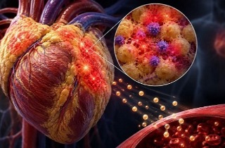
optical imaging technique that found differences between potentially life-threatening coronary plaques and those posing less imminent danger for patients with coronary artery disease. Their method may give cardiologists additional data to identify patients at higher risk of future
heart attacks and help them improve medical therapy.
.jpg)
Currently,
coronary artery disease is the most common type of heart disease in the United States and also globally. It is caused by deposits of atherosclerotic plaques within coronary arteries that supply blood to the heart muscle. Atherosclerotic plaques are accumulated, lipid-rich (fatty) material and calcifications in the vessel wall, and cause the inside of the arteries to stiffen and become restricted over time.
Typically, the most common symptom of
coronary artery disease is angina, which causes chest pain or discomfort during physical exertion because the plaques restrict blood flow and oxygen supply to the heart muscle. Also called stable angina pectoris, this condition may weaken heart function over time, but does not usually cause a sudden heart attack. But when the fibrous cap covering a lipid filled atherosclerotic plaque ruptures and releases the plaque content into the vessel, a blood clot can result that blocks the coronary artery and causes a
heart attack. This abrupt, life-threatening condition is called acute coronary syndrome. Cardiologists would like to be able distinguish stable coronary plaques from those prone to rupture.
Dr Behrouz Shabestari, Ph.D., director of the NIBIB program in Optical Imaging and Spectroscopy told
Thailand Medical News "This research relies on the latest optical imaging techniques to systematically categorize plaques as indicators of
coronary artery disease.The technique could be a game changer for cardiologists and their patients, offering refined insight into coronary arterial atherosclerotic lesions with quantifiable imaging data."
The study team is led by Dr Brett Bouma, Ph.D., professor of dermatology and health sciences and technology, Wellman Center for Photomedicine, Massachusetts General Hospital and Harvard Medical School, Boston. In a study published in the Aug. 8, 2019 issue of JACC: Cardiovascular Imaging, Bouma's team investigated the polarization properties of coronary atherosclerotic plaques in 30 patients with coronary artery disease, searching for indications of plaque instability.
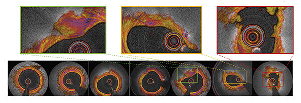 Optical imaging distinguished seven categories of coronary arteries, from left: normal, fibrous, fatty,
calcified, thick cap, thin cap, ruptured cap. Credit: Otsuka, K. et al. J Am Coll Cardiol Img
The researchers gathered data using intravascular polarimetry, a technique for assessing the polarization properties of c
ross-sections of vascular tissue. The electric field of polarized light creates a wave signal along a single plane (such as vertical or horizontal). When it is directed at tissue, the electric field is influenced by the microscopic structure and organization of the tissue. Tissues rich in collagen and smooth muscle cells split the beam of light into two rays that diverge into slightly different directions. The researchers could distinguish
coronary plaque composition and stability based on the optical effect.
Patients in this study underwent cardiac catheterization including intravascular imaging with optical coherence tomography, to measure the polarization properties of the coronary artery wall. Intravascular imaging uses light in the near infrared range to acquire high-definition, cross-sectional images of the vessel wall. Twelve catheterizations were performed on patients who had been affected by acute coronary syndrome the higher risk form of the disease, and another 18 on patients with symptoms of stable angina pectoris.
The thirty catheterizations provided multiple plaque images for each procedure, including 342 cross-sectional plaque images and 244 images from the fibrous caps of the atherosclerotic lesions responsible for high risk or stable symptoms. The high-resolution images enabled the researchers to classify coronary cross-sections into one of seven categories: normal, fibrous, fatty, calcified, thick cap, thin cap, or ruptured cap. Then, the team used the specialized instrument to determine the polarization properties of the coronary arterial wall.
Dr Brett Bouma further commented, "This is the first-in-human pilot study of intravascular polarimetry. Noting that fibrous caps of plaques that are prone to rupture can now be reliably identified using the method. Intravascular polarimetry may open new avenues for studying plaque composition and detecting high-risk patients."
Reference : Kenichiro Otsuka et al. Intravascular Polarimetry in Patients With Coronary Artery Disease, JACC: Cardiovascular Imaging (2019). DOI: 10.1016/j.jcmg.2019.06.015