

BREAKING NEWS
For the latest on Thailand Medical Industry, Thailand Doctors, Thailand Medical Research, Thailand Hospitals, Thailand Wellness Initiatives and the latest Medical News
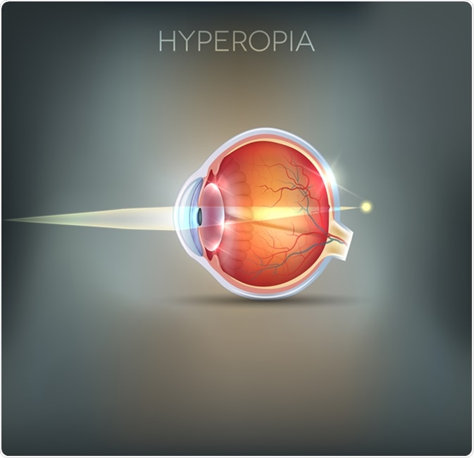
The ophthalmological condition that presents with deficiency in near vision, but normal far vision, is known as hyperopia or farsightedness.
While most term babies are born with this condition, it usually self-corrects by the age of 5. However, it may still be found in up to 10% of the adult population.
Hyperopia is more prevalent in persons of Native American and African American as well as Pacific Islander descent.
Persons who are young and have adequate reserve of accommodative function may not experience any symptoms. Those who are outside this age group, that is, the very young and very old populations, will experience problems.
Asthenopia, or eyestrain, is a frequent complaint, together with headaches, blurred vision, eye fatigue and occasional double vision. Other signs and symptoms include red eyes and increased tearing with decreased binocularity.
Farsightedness arises due to an error in the refractive properties of the eye, causing light to be focused at a point behind the retina. Those affected may have an eyeball that is too short in length, or pathologies in the lens or the shape of the cornea.
A comprehensive examination of the eye with a series of different tests may be used to diagnose hyperopia. During these tests, an ophthalmologist will examine the retina and conduct tests of muscle integrity, slit-lamp, refraction, visual field and visual acuity tests, which help to ascertain the condition. Examination of the retina is called ophthalmoscopy or funduscopy. An ophthalmoscope allows for retinal, optic disk and choroid evaluation. Muscle integrity is conducted by observing the eye movement responses to a moving object.
Refraction tests help to assess how well the cornea and lens bend the light. They also assist with prescribing the correct strength lenses. The slit-lamp is used to magnify and illuminate the frontal ocular area, which includes the lashes, eyelids, iris, cornea, lens and fluid chambers. Perimetry or testing of the visual field is necessary to identify possible defects, while visual acuity is tested with the help of characters of varying sizes on a chart at a standard distance away from the patient.
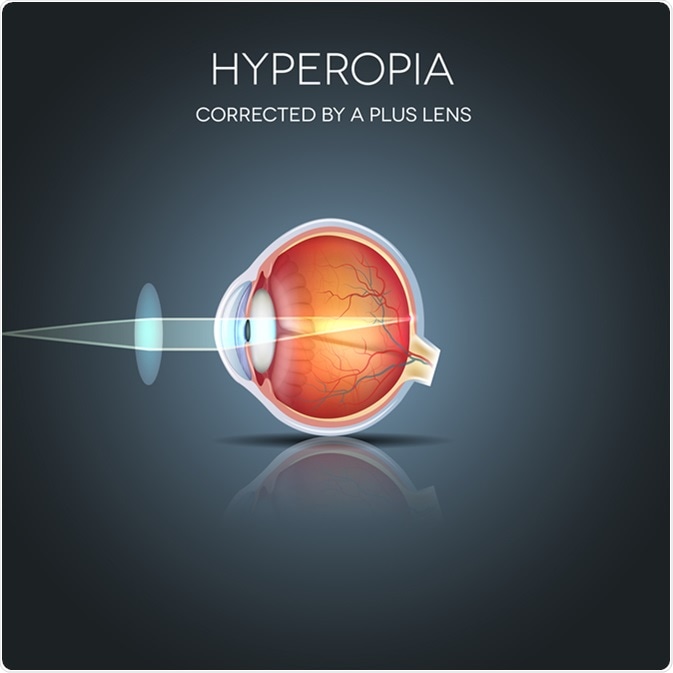
The most common means employed to treat hyperopia include eyeglasses and/ or contact lenses. These aids work by focusing the light rays onto the retina, which allows the development of a clear, normal image. Spherical lenses may be used to help achieve this. If there is a problem with the curvature of the cornea, then contact lenses may be used. Several types of lenses (e.g. soft, hard, gas permeable, extended-wear and disposable) are available. In addition to eyeglasses and contact lenses, refractive surgery may also be done to help correct hyperopia, even though this is mostly done for patients with short-sightedness.
There are several different types of refractive surgeries for hyperopia. In laser-assisted in-situ keratomileusis, one of the refractive surgical procedures, a flap cut is made into the cornea and a laser is used to increase the dome shape of the cornea. Another type of refractive surgery is laser-assisted subepithelial keratectomy, where no flap cut is made, but the outer epithelial layers of the cornea are re-shaped with a laser. These procedures, like all surgical procedures, are accompanied by potential risks and complications, which should be taken into account. These include, but are not limited to, over- and under-correction, infection, inflammation and pain.